


Femoral Neck & Hip Fracture Overview
Femoral Neck & Hip Fracture Overview
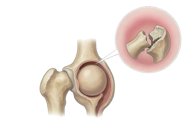
A hip fracture is a break in the upper part of the thigh bone, and the first thing that matters about it is where it sits. A break in the neck lies inside the fibrous sleeve that carries the blood supply to the ball, so a displaced fracture there can starve the ball of blood. A break in the trochanteric block or just below it lies outside that sleeve, in spongy bone that heals well. That one distinction, read with how far the fragments have moved and with what the patient could do before the fall, decides whether the fracture is fixed or the hip is replaced.
-
48 hours
Written opinion from our orthopaedic team once your images and records arrive complete
-
Day one
Sitting out of bed and starting to walk the day after surgery, where the surgeon permits
-
Two questions
Will the ball of the joint survive, and will the bone hold the implant
-
One team
Surgeon and geriatric physician review an older patient together, not in sequence
What a hip fracture is, and why its position decides the operation
The word hip is used loosely in everyday speech, and two things need separating at the start. The hip is a joint: the ball at the top of the thigh bone sitting in a socket in the pelvis. A hip fracture is not a break of the pelvis or of that socket. It is a break in the upper end of the thigh bone itself — the femur — in the part that carries the ball. That upper end has four parts: the head, the ball; the neck, which joins it to the rest of the bone at an angle; the trochanters, where the hip muscles attach; and the shaft below, which carries the length of the leg.
A fibrous sleeve called the capsule wraps the joint, attaching along the rim of the socket and coming down to the base of the neck. The head and neck sit inside it; the trochanters and shaft sit outside. Fractures inside that sleeve are intracapsular; those outside are extracapsular. Almost every femoral neck fracture is intracapsular, and almost every intertrochanteric or subtrochanteric fracture is extracapsular. The reason is one of plumbing. The head of the thigh bone has almost no blood supply from inside the bone; the vessels that keep it alive run up the neck within the capsule. A fracture that displaces the neck can tear or kink them, and a head that loses its supply dies — a process called avascular necrosis — so the fracture may not unite at all. The trochanteric and subtrochanteric zones are spongy bone wrapped in well-vascularised muscle, and they bleed when they break, which means they have a supply, and they heal.
Two completely different questions therefore come out of the same part of the body. For an intracapsular fracture the question is whether the head will survive, and the answer decides whether the fracture is pinned to keep it or the hip is replaced. For an extracapsular fracture the question is which implant will hold a broken block of spongy bone while it heals, and the fracture is almost always fixed rather than replaced. One more thing shapes everything. In an older adult this injury is not a painful inconvenience to be worked up at leisure: somebody who cannot take weight cannot be turned comfortably, does not drink enough, becomes confused, and is at risk of pressure injury, chest infection and blood clots. That is why the diagnosis is handled as an emergency — admit the same day, assess, and operate as soon as the patient is fit. Watching a hip fracture for a few days to see how it settles is not a safe option.
It helps to know what this condition is not. A worn hip joint that aches on walking, with no injury, is hip and knee osteoarthritis, not a break; a ball that dies without a fracture is avascular necrosis of the femoral head; and a collapsed vertebra in an osteoporotic spine is a vertebral fracture, a different disease.
Three zones in the upper thigh bone, and the one that changes everything
Hip fractures are named by which of three zones they pass through, and the zone sets how the blood supply is arranged, how the bone behaves when it breaks, how likely it is to heal, and which operation is even possible.
| Zone | Where the break runs | What it means in practice |
|---|---|---|
| Femoral neck | The strut between the ball and the trochanters, inside the capsule | The zone where the blood supply to the ball is at risk. Displacement is the central problem, and in an older patient the choice is often between fixing the fracture and replacing the head |
| Intertrochanteric | Across the block between and around the two trochanters, outside the capsule | The commonest hip fracture in older adults. Spongy bone with a good blood supply, so it heals; the question is which implant will hold it, and whether the outer wall is intact |
| Subtrochanteric | The first few centimetres of the shaft, below the lesser trochanter | High mechanical load with powerful muscles pulling across it. Usually held with a nail passed down the inside of the bone, and healing can be slower |
The distinction that decides the operation is not the zone but the capsule, which cuts across the anatomy. Inside it, the vessels that feed the ball run up the neck, so a displaced femoral neck fracture can interrupt them — the route to avascular necrosis and to a fracture that will not unite, and the reason the choice there is between keeping the ball and replacing it. Outside it, the blood supply arrives from the surrounding muscle and the bone is spongy rather than a thin strut: these fractures bleed, and because they bleed they heal, so the question is not whether the bone will live but which implant will hold it.
Two further labels sit alongside the zones. A hip fracture is traumatic when it follows a fall from a height in otherwise sound bone; fragility when it follows a fall from standing height or less in bone already weakened, which is the usual situation in an older adult; and pathological when something else weakened the bone, most importantly a tumour or an infection, which is why an unusual fracture with no injury to explain it is imaged thoroughly rather than assumed. Age then reorders the picture. In a younger adult with a displaced neck fracture the priority is to save the native ball, because the hip has to last decades and no artificial joint does; in an older adult it is standing as soon as possible, and the artificial joint is usually the better answer.
How severity is read: how far it has moved, and what the patient can bear
Describing a hip fracture as mild or severe is not useful, because two separate questions are being asked at once. The first is about the fragments: how far have they moved apart, and will the bone join again. The second is about the person: what was this hip being asked to do before the injury. The first is graded for neck fractures by the Garden classification.
| Grade | What has happened to the fragments | What it means |
|---|---|---|
| Garden I | Incomplete, or impacted with the neck tilted the other way. The break does not run right across the bone | The attachments carrying the blood supply are largely preserved. Some people can still walk on it, which is exactly why these fractures get missed |
| Garden II | Complete, but the fragments have not moved out of line | Blood supply largely intact. Grades I and II are called undisplaced, and are treated by fixing rather than replacing the joint, at almost any age |
| Garden III | Complete and partly displaced: the neck has tilted, fragments no longer in line though still touching | The vessels up the neck are stretched and compressed. The fracture may still unite, but the risk to the ball has risen, and age starts to change the recommendation |
| Garden IV | Complete and fully displaced: the head has separated and rotated back into the socket | Blood supply to the ball is most likely torn, so this grade is the least likely to unite and the most likely to be followed by avascular necrosis. In an older patient the head is usually replaced |
Extracapsular fractures are not graded with Garden at all. What matters there is stability: whether the broken block has an intact outer wall to push against, and whether the fracture line runs in a direction a screw-and-plate can hold.
| Pattern | What the fracture looks like | Why it matters |
|---|---|---|
| Stable | The break runs from the outer trochanter down to the inner one, and the outer wall is unbroken | The fragments can be compressed against one another. A screw that slides through a plate, letting the bone settle into a stable position as it heals, works well in many of these |
| Unstable | The outer wall is broken, the bone is fragmented, or the fracture line runs the opposite way | A screw and plate can lose its hold and the bone can collapse. These usually need a nail passed down the inside of the bone, which carries load closer to the axis of the leg |
| Extending below the trochanters | The break continues into the upper shaft, so one fracture becomes two patterns in one bone | A high-load region in which simple devices fail. The pattern is planned in detail before the operation, and a nail is almost always chosen |
The third axis is the patient, and in the neck it is often the axis that decides. What matters is how well the patient was walking beforehand, whether memory and thinking are intact, how much bone there is, and what the patient needs to be able to do afterwards. A person who was independent at home is treated differently from one who was already dependent in a care home, with identical images. The classification describes the fracture, and the patient decides the operation.
Finally there is time. A hip fracture has a window that closes: the sooner the bone is stabilised and the patient is up, the shorter the period of immobility and its consequences. Surgery is arranged as soon as the patient is medically fit, and within about 48 hours is the benchmark hospitals are measured against.
How a hip fracture is confirmed — and the fracture a plain X-ray can miss
Confirming a hip fracture is not usually difficult. A patient who has fallen, cannot get up, and has a leg that is short and turned outwards is straightforward, and a radiograph settles it. The interesting problems are at the edges: the patient who can still walk, the patient whose film looks normal, and the patient whose fracture pattern must be known precisely before an implant is chosen. The first images are an anteroposterior radiograph of the pelvis and a lateral view of the injured hip, and both are needed. The pelvis film shows both hips together, so a difference in the angle of the neck, or a step in the smooth curve where the neck meets the socket, is easier to see by comparison than alone. The lateral view shows the neck end-on, where an undisplaced fracture and any backward tilt of the head become visible.
What the surgeon reads from those two images is a short list: which zone the fracture is in; how far the fragments have moved, and therefore the Garden grade for a neck fracture; whether the bone is fragmented, and whether the break runs in a direction a screw will hold; whether the outer wall of the trochanteric block is intact; and whether anything else has broken, because a second fracture changes the plan. A bone-health assessment — a scan of bone density and blood tests for vitamin D, calcium, thyroid and parathyroid function — is completed during the admission rather than months later, because a hip fracture in an older adult is itself a statement about the skeleton.
| Investigation | The question it answers | What it cannot tell you |
|---|---|---|
| Pelvis and hip radiographs | Is there a fracture, which zone is it in, and how far have the fragments moved | The state of the blood supply to the ball, and often nothing at all about a fracture that has not displaced |
| CT of the hip | The exact pattern: fragmentation, the outer wall, and whether the break runs on below the trochanters, all of which choose the implant | It is not the most reliable test for a fracture that plain films have already missed, and it does not show the vitality of the ball |
| MRI of the hip | Whether there is a fracture at all, when the radiograph is normal but the hip is painful and will not take weight | It does not choose the implant, and it needs a scanner and a slot — it is not an imaging test every hospital can do at three in the morning |
The patient whose radiograph is normal is the one worth remembering, because it is where a hip fracture can be lost. When somebody who has fallen has a painful hip, cannot take weight, and cannot lift the leg straight against gentle resistance, the fracture is treated as present until it is proved otherwise, whatever the film shows. A proportion of undisplaced neck fractures are not visible on the first radiograph, and the bone can be walked on for days in that state, displacing as it goes. MRI is the most sensitive test; where it is not available, CT is used. Sending somebody home with a painful hip, an inability to weight-bear and a normal film is the error to avoid.
Why patients come to China for hip fracture care
The honest starting point is that a fresh hip fracture is not a condition to travel with. The bone needs to be stabilised where the patient is, as soon as they are fit, and flying with an unoperated hip fracture, or in the days after one, is dangerous. Where our network is genuinely useful is in what comes next, and in the cases that have already become complicated: a hip that was fixed and did not unite, a fracture around an existing replacement, a ball that lost its blood supply after a neck fracture, or an old fracture that has healed in a poor position and now needs the joint rebuilt.
The trauma and implant side
Complex hip work is a decision tree, and working through it in one place is what matters. Our orthopaedic teams hold the full range of devices for this region — screws through the neck, a screw that slides through a plate, nails passed down the inside of the thigh bone, and the complete range of artificial joints from a replacement of the ball alone to a full joint replacement. Revision work is part of the same service, so a fracture around an existing implant, or a hip that has failed to unite, is assessed by the team that can also rebuild it. Where the anatomy is unusual, the operation is planned in advance on a physical model printed from the scan data, produced in a 3D printing centre run jointly with the academician Dai Kerong team of Shanghai Ninth People's Hospital, and our orthopaedic robot is used where instrumentation must be placed precisely.
The frailty and bone side
A hip fracture in an older adult is not only an orthopaedic problem, and treating it as one leaves half of it undone. Every older patient is reviewed by a physician alongside the surgeon, with attention to heart and lung function, kidney function, the medicines that cause drowsiness, memory and thinking, nutrition and the risk of pressure injury — because these, and not the bone alone, decide how the first two weeks go. The admission then ends in a bone plan: the choice and timing of treatment for osteoporosis, a falls assessment with specific changes written down, and a follow-up schedule with the imaging intervals built into it. Patients continue through a structured rehabilitation programme, with the later stages completed at home.
The practical side matters too, particularly for a patient already in China or able to plan ahead. Records and images are reviewed by an orthopaedic surgeon working with the geriatric team, and a written opinion is returned within about 48 hours of complete records arriving, setting out what the images show, which available operation fits the fracture and the patient, and what the bone assessment should include. Our network runs 18 hospitals, works with more than 30 commercial insurers for direct billing where cover allows, and has received patients from 124 countries and regions.










