


Osteoporotic Vertebral Fracture Overview
Osteoporotic Vertebral Fracture Overview
An osteoporotic vertebral fracture is the collapse of a vertebral body made weak by bone loss. It behaves unlike the other conditions in this section: the pain is not brought on by walking or bending so much as by load — turning in bed, rising from a chair, standing — and it often begins after an event so minor that nobody thought of it as an injury. Two questions shape everything that follows: how much of the vertebral body has collapsed, and whether the fracture is still fresh.
-
30,000+
Spine operations performed by the spinal orthopaedic team
-
48 hours
Written opinion after complete records arrive
-
4 platforms
Microscope, microendoscopic, transforaminal and interlaminar endoscopy
-
9 stages
Structured post-operative rehabilitation assessment
What an osteoporotic vertebral fracture actually is
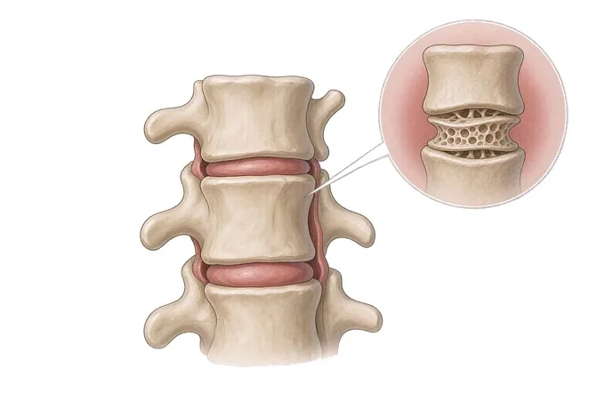
A vertebral body is a block of bone with a honeycomb structure inside: a fine lattice of struts within a thin shell, strong the way a cardboard box is strong — because of how it is built rather than how much material it contains. Osteoporosis thins those struts and the shell. What is left still carries ordinary load, but it has lost the reserve that a fall, a heavy lift or a sudden movement draws on. An osteoporotic vertebral fracture is what happens when that reserve runs out: the front of the body collapses, the block becomes a wedge, and the spine changes shape at that level.
The pain this produces does not behave like the pain of a pulled muscle or an arthritic spine. It is a load-dependent pain. Whether you are standing, sitting, walking or lying down matters more than what you did, because the fractured body is at its most comfortable when it is not carrying anything. Patients describe turning over in bed, getting out of a chair and getting up out of a low car seat as the worst moments of the day — and lying flat as the reliable relief. That pattern is unusual enough among back pains that it is worth taking seriously on its own.
Two things then follow, and they are the reason this condition is treated as more than a painful episode. The first is shape: with each fracture the spine loses height and the upper back becomes more rounded, which changes balance and shortens the trunk. The second is risk: a vertebra that has already fractured once is a statement about bone strength, and a further fracture becomes considerably more likely than the first one was. A vertebral fracture is therefore both a painful event and a piece of information about the skeleton that should change what happens next.
One caution belongs near the beginning. A compressed vertebra on a scan is not automatically the cause of the pain being reported: deformities are common in older adults and many are old. Fresh and broken are separate questions, and deciding which level explains which symptom is the judgement that determines whether any procedure is worth doing.
It is also worth distinguishing this from the other lumbar conditions that share its symptoms. A herniated lumbar disc produces pain in a band down one leg and is worst when sitting. Lumbar canal narrowing produces a walking limit in both legs. Spondylolisthesis is a segment that has slipped and moves. None of them is caused by loss of bone strength, and none of them is treated by treating the bone — which is the subject of this page.
Four fracture shapes, and what each one means
Vertebral fractures are described by the shape the vertebral body takes, and the shape is not cosmetic — it reflects how the bone failed, whether the back wall of the vertebra is involved, and how likely it is that the spine will bend further. Four patterns are recognised.
| Shape | What has happened | What it means in practice |
|---|---|---|
| Wedge | The front of the vertebral body has lost height while the back has kept it, so the body becomes wedge-shaped | The commonest pattern, and the one that produces the forward curve of the upper back. It is also the pattern most often found by chance on a chest or spine film taken for another reason |
| Biconcave | Both endplates have dipped inwards so the body becomes concave above and below, while the front and back walls keep their height | Associated with softening of the bone and with disc material pressing into a weakened endplate, and often less painful than the other patterns |
| Crush | The vertebral body loses height throughout — front, back and both endplates | The pattern with the greatest loss of height and the greatest effect on the shape of the spine, and often the most painful in the acute phase |
| Burst, with the back wall involved | The fracture extends into the back wall of the vertebral body, and a fragment may be pushed towards the canal | The pattern that decides against simple cement injection: the back wall must be intact before cement is placed, and if the cord or nerves are compressed the plan changes to decompression and stabilisation |
The practical value of the distinction lies in the fourth row. Almost every other feature of a vertebral fracture can be discussed in terms of pain and comfort, but the state of the back wall determines the safety of the techniques most often asked about — which is why imaging and examination are put together before an operation is proposed, rather than after.
Two further labels are used alongside the shapes, and one of them changes the whole plan. A fracture is traumatic after a major injury, osteoporotic after minor or no injury in weak bone, and pathological when the cause is something other than bone loss — most importantly tumour or infection. The last of these is why a first episode of this kind is imaged rather than assumed.
How severity is graded: how much height was lost, and whether it is fresh
Two questions are asked at the same time when a vertebral fracture is assessed, and they are independent of each other. The first is how much of the vertebral body has collapsed. The second is whether the fracture is still recent. A severe fracture that happened four years ago and a mild fracture that happened last week are, for practical purposes, at opposite ends of the spectrum.
The first axis is usually described with the Genant semi-quantitative grade, which classifies the shape of each vertebra from normal to severely deformed. It is a description of the architecture rather than a measure of the pain, which is why it is used together with what the patient reports.
| Grade | Height lost | How it is described in practice |
|---|---|---|
| Grade 0 | Up to about a fifth | The vertebra is within normal limits for its level. Many vertebrae that look mildly reduced on a first glance fall here |
| Grade 1 — mild | Around a quarter | A deformity that is clearly present when the level is compared with the vertebrae above and below, but not the level's dominant feature |
| Grade 2 — moderate | Around a third to two fifths | The level is obviously deformed. This is where height loss begins to be visible as a change in posture and in the length of the trunk |
| Grade 3 — severe | More than two fifths | The level is markedly collapsed and the local curve of the spine is affected. The back wall is examined with particular care at this grade |
The second axis is freshness, and it is the one patients most often arrive without having had assessed. A fracture still healing behaves differently from one that has healed, and the treatments most asked about — cement injection among them — apply mainly to the first situation. Freshness is established from the history together with imaging that shows oedema inside the bone, the signature of an active fracture.
A third axis overlaps both, and it is function rather than anatomy: the amount of pain and the degree of movement it allows. Two people with an identical grade, at an identical stage of healing, may differ entirely in what they can do. It is this axis that determines how urgently something needs doing, and it cannot be read from any image.
What overrides both of these axes is a neurological deficit: progressive weakness in the legs, or a change in bladder or bowel control. Where these are present, the conversation is no longer about pain management but about assessing the level and degree of neural compression the same day.
How a vertebral fracture is confirmed and dated
The assessment of a suspected vertebral fracture answers three questions, and they are asked in a specific order: is there a fracture, how recent is it, and what caused it. Skipping to the third question is a common mistake, and so is treating the first as if it settled the other two.
Radiographs of the thoracic and lumbar spine are the first step, taken from the side as well as from the front, because a compression fracture is invisible on a frontal film alone. The films are read with the vertebrae above and below as the reference. Where the question is whether the fracture is still active, MRI adds what the radiograph cannot: it shows oedema inside the vertebral body, present while a fracture is healing and gone once it has healed, and it shows compression of the cord or nerves where the back wall is involved.
Three investigations then divide the work between them, and it is worth knowing which question each one answers, because patients frequently arrive having had one of them and assuming the other two are settled.
| Investigation | The question it answers | What it cannot tell you |
|---|---|---|
| Lateral radiographs | Whether there is a fracture, and what shape the vertebral body has taken | Whether the fracture is recent, or what the nerves are doing |
| MRI, and CT where needed | Whether the fracture is still healing, and whether the canal or a nerve is compressed | How strong the bone is elsewhere, or what is causing the loss |
| Bone density and blood tests | How much bone has been lost, and which correctable factors are contributing | Whether this particular vertebra is fractured, or whether it hurts |
The last of these is measured rather than assumed, and it is not a way of diagnosing the fracture: a density result describes the skeleton, not the injury. Blood tests complete the picture by looking for correctable contributors — vitamin D deficiency, parathyroid and thyroid problems, markers of bone turnover — and by looking for the causes that are not osteoporosis at all.
That last point is the reason this assessment is not simply a formality. Tumour and infection both produce a collapsed vertebral body, and both can be mistaken for an osteoporotic fracture on plain films. The features that separate them are read from the history, the examination, the MRI and the blood tests together.
Why patients travel to China for vertebral fracture treatment
The reason is not that the procedure is exotic — cement injection has been performed for decades. It is that this condition sits between two specialities, and the decision that matters is which one the case belongs to: bone medicine, or spine surgery. A useful assessment has to be able to answer both, and that is what an integrated spine service offers.
The procedural side
Vertebroplasty and kyphoplasty are performed under imaging guidance by surgeons who also perform decompression and fusion, so the alternative operations are genuinely on the table. Where the back wall of the vertebra is broken, or the cord or nerves are compressed, the plan is changed before the procedure rather than after it. The department works through four endoscopic and microscopic platforms and uses an orthopaedic robot covering the cervical, thoracic, lumbar and sacral spine where instrumentation is required, with each case planned through 26 standardised nodes. Complex and revision cases are rehearsed on physical models from a 3D printing centre run jointly with the academician Dai Kerong team of Shanghai Ninth People's Hospital.
The bone and safety side
Treating the painful level without treating the skeleton is an incomplete treatment, because a further fracture is the next thing a patient is at risk of. Our assessment therefore ends in a bone-health plan rather than in a procedure alone, including the choice of osteoporosis medication and its timing around any intervention, a falls assessment with specific advice, and a follow-up schedule with the imaging intervals written into it. Patients are followed through a structured nine-stage rehabilitation assessment, and the later stages are completed with the patient's own doctor at home.
The practical side matters as much as the clinical one. Records are reviewed by a spinal surgeon working with the bone health team, and a written opinion is returned within about 48 hours of complete records arriving. We work with more than 30 commercial insurers for direct billing and have received patients from 124 countries and regions. It is worth knowing in advance that a review can conclude that no procedure is indicated — and that outcome is written down as clearly as any other.










