


Lumbar Disc Herniation Overview
Lumbar Disc Herniation Overview
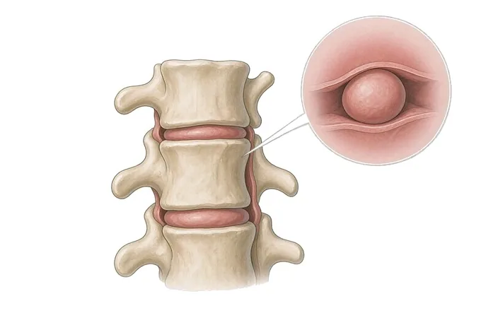
A herniated lumbar disc is a tear in the outer ring of a disc through which the softer inner material pushes — usually backwards and to one side, which is why the pain runs down a leg rather than staying in the back. Most people improve without an operation, and the two things that decide the plan are how far the disc has come out and how the nerve is behaving.
-
30,000+
Spine operations performed by the spinal orthopaedic team
-
4 platforms
Microscope, microendoscopic, transforaminal and interlaminar endoscopy
-
48 hours
Written opinion after complete records arrive
-
9 stages
Structured post-operative rehabilitation assessment
What a herniated lumbar disc actually is
Each lumbar disc has two parts. Around the outside is the annulus fibrosus, a ring of tough layered fibres; in the centre is the nucleus pulposus, a gel that holds water and behaves like a shock absorber. A herniation begins as a tear in the fibres of the outer ring. Once the tear reaches far enough, the gel is pushed out along it — and because the back of the disc sits directly against the nerve roots that run down the leg, that is usually where the trouble starts.
The word slipped is the reason so much of this is misunderstood. A disc does not slide out of position: it stays between the two vertebrae, and it is the material inside it that escapes. That distinction matters practically, because it means the problem is usually a pressure and chemical irritation of one nerve root, not a spine that has moved and needs to be put back.
Almost all symptomatic herniations occur at one of the two lowest mobile levels, L4/L5 and L5/S1, and the nerve involved decides which part of the leg complains. That is why the examination is about myotomes, reflexes and the distribution of numbness rather than about the back itself: the back is where the problem is, but the leg is where it shows.
It is also worth knowing what a herniation is not. It is not the same as the ordinary age-related disc degeneration that nearly every adult has on a scan, and it is not the same as a bulge, in which the whole ring widens without a tear. A bulge is a shape; a herniation is a displacement of material. Whether the pattern in your case needs treatment at all is covered under symptoms and warning signs.
Four patterns of herniation, and why the pattern decides the plan
Herniations are described by the direction in which the material travels and by how much of the ring has failed. The direction decides which structure is compressed, and that in turn decides the symptoms and the operation — if one is needed.
| Pattern | Where the material goes | What it typically produces |
|---|---|---|
| Posterolateral (paracentral) | Backwards and to one side, into the space where the nerve root is leaving the canal | The commonest pattern: leg pain in a band, with a positive straight leg raise |
| Foraminal | Outwards into the opening through which the nerve exits | More intense pain in a narrower band, often with numbness rather than weakness early on |
| Central | Straight backwards into the canal | Back pain, and where the canal is narrow, symptoms in both legs or difficulty walking |
| Sequestered (migrated) | A fragment separates and moves up or down inside the canal | A more severe course in the first weeks, and an important reason to image before planning |
Two further terms appear on reports and are frequently confused with a herniation. A protrusion means the material is still contained by the outer fibres; an extrusion means it has broken through them; a sequestration means a free fragment. And an annular tear is a tear in the ring without displacement — it can be painful, but it is not the same diagnosis and is not treated with the same operation.
How a herniation is graded: two axes, not one
How far a disc has come out is only half the question, and on its own it is a poor guide to what should be done. What matters equally is how the nerve is behaving: a large herniation pressing on a nerve that is only irritated behaves very differently from a modest one pressing on a nerve that is losing power. The plan is read from both axes together.
| Axis 1 — the disc | Axis 2 — the nerve | What it usually means |
|---|---|---|
| Bulge (no tear) | No nerve symptoms | A shape on a scan, not a diagnosis: no treatment directed at the disc |
| Protrusion | Pain only, in a single band | The most common presentation; non-surgical treatment is the first step |
| Protrusion or extrusion | Sensory change, normal power | Still usually treated without surgery, with the neurological examination repeated at intervals |
| Extrusion | Measurable weakness or a lost reflex | The decision turns on whether the weakness is improving, stable or worsening |
| Extrusion or sequestration | Weakness that is progressing over days | Surgery is generally advised rather than watched, because recovery of power is time-dependent |
| Central or sequestered | Saddle numbness, urinary retention, bilateral symptoms | Cauda equina syndrome — an emergency, and the one situation with no waiting period |
The practical consequence of the second axis is a rule about time. Nerve pain can be given weeks because it responds to treatment and usually settles. Lost power and disturbed bladder function cannot be given weeks, because the longer a compressed nerve is left without function, the less of it returns. This is the single distinction that decides who should be assessed urgently rather than at the next convenient appointment.
How the back and the leg are assessed
An assessment for sciatica answers three questions in order: is the pain coming from a nerve root at all, which root is it, and is that nerve merely irritated or actually losing function. Everything else — including the scan — is there to confirm or refute those answers.
The distribution of the pain, what makes it worse, and the reflex and power findings, taken together, localise the level in most patients before any imaging. Straight leg raise and its crossed variant are the two most informative bedside tests, and they are repeated at intervals rather than performed once.
It shows the disc, the canal and the nerve together, which no other single test does. It is requested when symptoms persist despite a fair period of treatment, when a neurological deficit is present, or when the presentation is atypical — not as a routine first step, because most adults have disc changes that have nothing to do with their pain.
A standing radiograph is used when instability, deformity or a stress fracture is suspected, and it is essential before any fusion is planned. CT is useful where MRI cannot be performed, and for bone detail around a previous fusion.
Nerve conduction studies and electromyography separate a compressed nerve root from a problem further down the leg, such as a neuropathy or a trapped nerve at the knee or ankle. They are not routine, but they change management when the examination and the scan point in different directions.
What follows the assessment should be a written plan with dates in it: what is being treated now, what would trigger a change, and when the examination will be repeated. Details of each test and what it can and cannot show are in how a herniated disc is diagnosed.
Why patients travel to China for disc surgery
For a herniated disc the clinical decision is not usually the difficult part — the difficulty is finding somewhere that will make the decision carefully, offer the full range of options including the least invasive one, and do so without a waiting list long enough to make the decision for you.
The full range of access, not one technique
The spinal orthopaedic team works through all four minimally invasive platforms — microscope, microendoscopic, transforaminal endoscopic and interlaminar endoscopic — which matters because the right approach is decided by where the fragment sits and how the canal is shaped, not by which instrument a centre happens to own.
Sub-specialty allocation rather than a general list
Cases are allocated to a spinal surgeon whose routine practice is the relevant level and approach. For a herniation that is occasionally a decision between a 7-millimetre endoscopic route and a small open one, and it is made by someone who does both.
Planning where the anatomy is difficult
The hospital's 3D printing centre, established with the team of Professor Dai Kerong of Shanghai Ninth People's Hospital, prints and studies complex anatomy before an operation — of most value in recurrent herniation and in deformity, where the normal landmarks have changed.
Robotic navigation across all spinal segments
Where instrumentation is needed, screw placement is planned and navigated robotically from the cervical spine to the sacrum, and every case is worked through 26 standardised technical steps with a nine-stage post-operative assessment.
The scale behind this is a spinal orthopaedic service that has performed more than 30,000 spine operations and now runs over 3,500 high-complexity procedures a year, with a nursing and rehabilitation team of 106 and 200 supervised pre-operative case observations for trainees. International patients from 124 countries and regions are supported by direct billing with more than 30 insurers, and the hospital was the first in Foshan designated under the Guangdong–Hong Kong–Macao medicine and device connect scheme. A written opinion follows within 48 hours of complete records arriving.










