


Lumbar Spondylolisthesis Overview
Lumbar Spondylolisthesis Overview
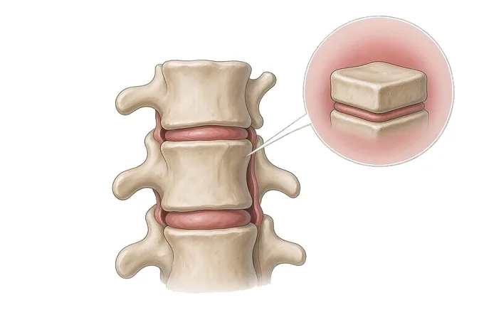
Lumbar spondylolisthesis means one vertebra has slid forward over the one beneath it, most often at the fourth or fifth lumbar level. It does two things at once: it takes space away from the nerves, and it introduces movement at a segment that is meant to be stable. Those two consequences explain why the same grade of slip can produce nothing at all in one person and disabling pain in another — and why the question that decides treatment is not only how far it has slipped, but whether it is still moving.
-
30,000+
Spine operations performed by the spinal orthopaedic team
-
4 platforms
Microscope, microendoscopic, transforaminal and interlaminar endoscopy
-
48 hours
Written opinion after complete records arrive
-
9 stages
Structured post-operative rehabilitation assessment
What a slipped vertebra actually is
The lumbar spine is a column of vertebrae stacked in a gentle curve, each one held to its neighbour by a disc in front and by two facet joints behind. Those joints guide movement and, just as importantly, stop one vertebra from sliding forward on the next. Spondylolisthesis is what happens when that restraint fails and a vertebra translates forward — the word itself comes from the Greek for a slipped vertebra.
There are two consequences, and they behave differently. The first is simple crowding: the vertebra that has moved takes space from the canal and narrows the opening through which the nerve root leaves, so the nerve is compressed in its tunnel rather than in the open canal. The second is mechanical: a segment that slides is a segment that moves abnormally, and that movement loads the disc, the joints and the muscles around it. Pain in spondylolisthesis usually comes from this second consequence; leg symptoms usually come from the first.
That is why the size of the slip on its own is a poor guide to how much trouble it causes. Some people have a substantial slip that has been stable for decades and produces nothing beyond occasional stiffness. Others have a modest slip that moves several millimetres every time they stand up, and they are limited every day. The grade describes the position; the movement describes the problem.
It follows that finding a slip is not the same as finding the cause of someone's pain. Slips are common in people over fifty, and a large proportion are incidental — present on a scan taken for another reason, and not the source of the symptom. Deciding whether a given slip is responsible means matching its level and its behaviour to what the patient actually describes, which is a clinical judgement and not a measurement.
One feature of that judgement is worth stating plainly at the outset, because it shapes everything that follows. In spinal stenosis without a slip, the symptom is typically a walking limit in both legs. In spondylolisthesis, the nerve is often compressed where it leaves the spine rather than in the middle of the canal, so the symptom is frequently one-sided leg pain in a band — closer to the pattern of a herniated lumbar disc than to the classic picture of lumbar canal narrowing. Getting that distinction right is the first step in reading the scan correctly.
Six types of slip — because the type is the cause
Spondylolisthesis is classified by what allowed the vertebra to move, and the classification is worth knowing because the type determines the age at which it appears, the level it affects and the operation that suits it. The traditional system describes six types.
| Type | What has failed | Who it affects, and where |
|---|---|---|
| Isthmic (pars defect) | A stress fracture through the pars interarticularis, the narrow bridge of bone between the upper and lower facet joints | The most common type in adolescents and young adults; typically L5/S1; strongly associated with sport involving repeated extension and rotation |
| Degenerative | Long-standing disc height loss and facet joint arthritis, which allow the segment to translate because the joints no longer interlock | The most common type overall; women over 50; almost always L4/L5; frequently accompanied by central canal narrowing |
| Dysplastic (congenital) | An abnormally shaped vertebra or facet joint present from birth, which never provided normal restraint | Presents earlier, sometimes in childhood; can reach high grades and may be associated with other spinal malformations |
| Traumatic | An acute fracture of the pars or of the facet complex from a single injury | Uncommon; follows identifiable trauma; the history and the imaging are usually decisive |
| Pathological | Bone weakened by disease — a tumour, an infection, or a generalised bone disorder | Uncommon, and managed by treating the underlying condition as well as the spine |
| Post-surgical (iatrogenic) | Removal of supporting structures at a previous operation, or degeneration at a segment next to a previous fusion | Appears years after lumbar surgery; a reason why preserving the facet joints during decompression matters |
Alongside the type, three measurements from the imaging carry most of the weight. The Meyerding grade describes how far the vertebra has slipped as a percentage of the vertebra below. The slip angle describes how far the slipped vertebra has tilted forward, which matters more than the translation in higher grades. And the condition of the disc at the slipped level — its height and whether it is degenerate — largely determines what the segment will do in the future, and whether a young patient with a pars defect is a candidate for repair rather than fusion.
Two situations are worth separating from the list. Spondylolysis is a pars defect without any slip; it is the stage that precedes isthmic spondylolisthesis, it is common in adolescent athletes, and it is treated differently — rest and graded return to sport, sometimes with a period of bracing. And a slip found by chance in someone whose pain has a different explanation is not a diagnosis; treating it as one is the most common error made with this condition.
How a slip is graded: how far it has moved, and whether it still moves
There are two questions, and only one of them appears in the radiology report as a grade. The first is anatomical: how far the vertebra has translated, how much it has tilted, and which level is involved. The second is mechanical and clinical: whether the segment still moves, how much of the pain is in the back rather than the leg, what the walking tolerance is, and whether the nerves are losing function. Treatment is read from the pair.
| Axis 1 — the slip | Axis 2 — movement and function | What it usually means |
|---|---|---|
| Meyerding I (under 25%), single level, no motion on flexion-extension views | Back pain that is manageable, no leg pain, full walking tolerance, no neurological findings | Observation and a strengthening programme: the slip is real but is not the limiting problem |
| Meyerding I–II with a normal or near-normal disc | Episodic back pain related to sport, hamstrings tight but power and sensation intact | Structured non-surgical treatment first; in young patients with an isthmic defect, pars repair may be an option later |
| Meyerding II, one-sided foraminal narrowing | Leg pain in a band on one side, worse standing and walking, eased by sitting | Injection and physiotherapy first; decompression considered if the pain persists despite adequate treatment |
| Any grade with abnormal motion on flexion-extension views | Back-dominant pain, a segment that feels unstable, symptoms on changing position | This is the finding that makes fusion part of the operation rather than decompression alone |
| Meyerding III or above, or a markedly increased slip angle | Difficulty standing upright, altered gait, tight hamstrings, walking limited | Planned reduction and instrumented fusion, assessed in detail before surgery rather than during it |
| Any grade with a neurological deficit | Measurable weakness, a foot drop, or disturbance of bladder or bowel function | Assessed promptly rather than observed; a high-grade slip can compress the cauda equina |
Read as a pair, these two axes explain the decision patients find most surprising: a larger slip is not automatically a stronger reason to operate, and a smaller slip is not automatically a reason not to. A stable Meyerding II slip with leg-dominant pain and a normal examination may be treated for years without an operation. A Meyerding I slip that translates visibly on standing films and produces back-dominant pain with every change of position may be the one that needs the segment addressed surgically.
The one element that overrides both axes is neurological deficit. A slip alone, however large, does not usually remove power; when power, sensation or bladder function are declining, the timescale changes from months to days, and that situation is covered under urgent assessment.
How a slip is measured and what has to be done standing
Because a slip is a position, the way the images are taken changes what they show. This is the single most practical point on this page, and it is the reason a report from a scan taken lying down may understate the problem.
The radiograph has to be standing
A slip is load-dependent. Lying flat, with the spine unloaded, a vertebra that translates several millimetres when you stand may look almost normally aligned. Standing lateral radiographs are therefore the baseline, and flexion and extension views are added because the difference between the two positions is what shows whether the segment is mobile.
MRI is for the nerve and the disc
MRI shows what the slip is doing to the nerves, whether the compression is central or in the exit opening, and the state of the disc at the slipped level — which is the measurement that most affects planning. It is taken lying down, which is precisely why it answers a different question from the standing films rather than replacing them.
The examination contributes findings that no image provides: whether there is a palpable step, how the patient walks, whether the hamstrings are tight, whether power and reflexes are intact, and how the segment behaves when the spine is extended on one leg. In adolescents with back pain after sport, that last test — the single-leg hyperextension or stork test — is often the finding that prompts the correct imaging.
CT is used when a pars defect is suspected and the plain films are inconclusive, because it shows the bony bridge directly. In an adolescent with a fresh stress injury, a bone scan or SPECT can distinguish an active lesion, which may heal with rest, from an old healed defect, which will not. Long cassette or whole-spine radiographs are added only when overall alignment or a deformity is part of the picture.
Why patients travel to China for spondylolisthesis surgery
The operation for a slipped vertebra is well established. What a centre adds is judgement about which of three quite different operations is right — decompression alone, decompression with fusion, or repair of the defect without fusing the segment — and the discipline to say when none of them is needed.
That judgement has a technical basis. Decompression alone is smaller, preserves movement and is appropriate for a low-grade slip that does not move. Fusion stabilises the segment but adds implants, a longer recovery and its own long-term considerations, so it should be used because the segment is unstable and not because a slip is visible. And in a young patient with an isthmic defect and a healthy disc, pars repair — fixing the fracture so the segment keeps its movement — can be preferable to fusion, provided the disc is good enough to justify it. Whether that applies is decided from the films, not from the patient's age alone.
Where fusion or reduction is required, planning is where the risk sits. Our spinal orthopaedic team has performed more than 30,000 spine operations and carries out around 3,500 complex (level-4) procedures a year, with 106 clinicians. Each case passes through 26 standardised planning nodes, including the levels to be fused, the degree of reduction to attempt and the trajectory of every screw. Stereotactic navigation and an orthopaedic robot covering the cervical, thoracic, lumbar and sacral spine are used for screw placement, and decompression is performed through four endoscopic and microscopic platforms — a microscope, a microendoscopic system, and transforaminal and interlaminar endoscopes — matched to the level and to the pattern of compression.
Since 2020 the department has run a 3D printing centre jointly with the academician Dai Kerong team of Shanghai Ninth People's Hospital, used to rehearse high-grade and revision cases before surgery, and each surgeon observes around 200 cases in theatre before operating independently.
For international patients the practical difference is how much can be settled before travelling. Send your standing and flexion-extension films together with the MRI, and a spinal surgeon will return a written opinion within 48 hours of complete records arriving, stating whether the segment is unstable, whether fusion is required, and how many levels would be involved. We treat patients from 124 countries, work directly with more than 30 commercial insurers for direct billing, and hold 52 approvals under the Foshan drug and device access scheme. Recovery is assessed through a structured nine-stage programme rather than a single follow-up visit.










