


Lumbar Spinal Stenosis Overview
Lumbar Spinal Stenosis Overview
Lumbar spinal stenosis is a gradual narrowing of the tunnel that carries the nerves to the legs. It produces a very particular complaint — legs that ache, feel heavy or go numb after a fairly predictable walking distance, and settle within minutes of sitting or leaning forward. Narrowing is common with age; whether it needs an operation depends far more on how far you can walk than on the words used to describe your scan.
-
30,000+
Spine operations performed by the spinal orthopaedic team
-
4 platforms
Microscope, microendoscopic, transforaminal and interlaminar endoscopy
-
48 hours
Written opinion after complete records arrive
-
9 stages
Structured post-operative rehabilitation assessment
What lumbar spinal stenosis actually is
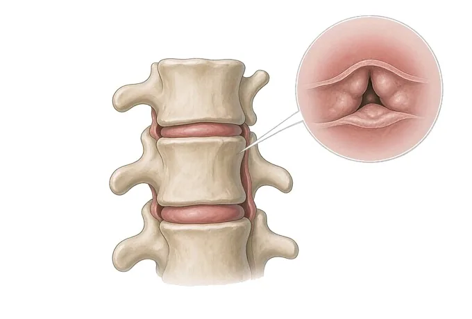
The spinal canal is a tunnel built from stacked vertebrae: discs at the front, pedicles at the sides, and the laminae and yellow ligament (ligamentum flavum) forming the roof. The nerves that serve the legs run down this tunnel and leave it through openings on each side. Stenosis simply means the tunnel has become narrower than it should be.
In the lumbar spine that narrowing is almost always the sum of three slow processes rather than one event. Discs lose water and height and bulge backwards into the canal; the facet joints at the back develop arthritis and enlarge; and the ligamentum flavum thickens and buckles inwards. Any one of these is tolerable — together, over decades, they take away the room the nerves need.
The condition is therefore cumulative and positional, not sudden: the canal in a 70-year-old has usually been narrowing for years and has simply crossed a threshold. Symptoms follow space and position rather than how much something hurts at rest. Standing and walking bring the nerve roots closer to the structures that crowd them and raise the demand on their blood supply; sitting or bending forward does the opposite.
It is worth separating this from a herniated lumbar disc, because the two are frequently confused and behave quite differently. A herniation is a discrete event affecting one nerve root and one leg, with pain in a band. Stenosis is a slow narrowing affecting several levels and both legs, with a limit on how far you can go rather than a pain that shoots. Where a herniated disc often hurts most on sitting, stenosis is generally worst on standing and walking.
One fact changes how patients read their own scan: narrowing is extremely common in older adults, and many people with clearly narrowed canals report no leg symptoms at all. The measurement that decides treatment cannot be seen on a scan — it is how far you can walk, and whether that distance is holding steady or shrinking.
Four places a lumbar canal narrows — and why the place decides the symptoms
Stenosis is described by where in the canal the narrowing sits, because each zone contains different structures and produces a different complaint. The terms on a radiology report refer to these zones.
| Pattern | Where the narrowing sits | What it typically produces |
|---|---|---|
| Central canal stenosis | The middle of the tunnel, from disc bulging, facet enlargement and inward folding of the yellow ligament | The classic picture: both legs ache, feel heavy or go numb after a set distance, relieved by sitting or leaning forward |
| Lateral recess stenosis | The side channel where the nerve root begins its journey outwards, before it reaches the foramen | More one-sided and more radicular: pain or numbness in a band down one leg, often with a positional element |
| Foraminal stenosis | The exit opening itself, narrowed by a collapsed disc, an enlarged facet or a bone spur | Constant one-sided leg pain, worse on standing and walking, often relieved by sitting and by flexing the spine |
| Multilevel stenosis | Two or more segments, often with a degenerated disc between them | A shorter walking distance and a more intrusive effect on daily life; surgery, when needed, must plan for every involved level |
| Developmentally narrow canal | No single zone of narrowing: the canal has been small since birth, usually because the pedicles are short | Symptoms decades earlier than usual, often at several levels and before much degeneration is visible; less reserve to lose |
Two modifiers appear alongside these patterns and change the operation more than the pattern itself. Degenerative spondylolisthesis means one vertebra has slipped forward on the one below, which both narrows the canal and introduces movement — the reason a decompression sometimes needs to be combined with a fusion. A synovial cyst arising from a facet joint can also occupy the canal and behave like an extra piece of tissue pressing on the nerve.
Radiologists grade central narrowing on MRI, most commonly with a four-point scale from A to D, where A describes a canal with visible fluid around the nerves and D describes one in which the nerves are pressed together with no fluid visible at all. The grade is useful for describing what is there and for comparing one scan with another. It is not a treatment plan, and treating the grade rather than the patient is the single most common mistake made with this condition.
How stenosis is graded: how narrow it is, and how far you can walk
There are two questions, and only one of them will be visible on your images. The first is anatomical: where the canal is narrow and how narrow it has become. The second is functional: how far you can walk before the symptoms force you to stop, how long it takes to recover, and whether that distance is stable or falling. The plan is read from the second axis, with the first used to decide whether an operation is technically feasible and which levels it should cover.
| Axis 1 — the canal | Axis 2 — walking and function | What it usually means |
|---|---|---|
| Narrowing present, fluid still visible around the nerves | Symptoms occasional, pain-free walking distance over about 500 metres, no unsteadiness | Observation and a walking programme: the anatomy is real but is not limiting your life |
| Moderate narrowing at one or two levels | Pain-free distance around 200 to 500 metres, needing to sit for a minute or two, distance roughly stable | Structured non-surgical treatment first — physiotherapy, medication, injection — with surgery if it stops working |
| Marked narrowing, nerves crowded together | Pain-free distance under about 200 metres, or stops and starts indoors; shopping or travel becoming difficult | Decompression is discussed seriously: the limitation is now the reason the operation exists |
| Narrowing with instability, slip or a cyst | Distance shrinking over months, or any new weakness, foot drop or unsteadiness | Decompression planned in detail, with fusion considered where there is movement at the level |
| Narrowing with a neurological deficit | Measurable weakness, a foot drop, or progressive unsteadiness rather than only a walking limit | Assessed promptly rather than observed: a nerve is losing function, and recovery depends on how long the compression lasts |
Read as a pair, these two axes explain a situation patients find puzzling. Someone whose scan is described as severe but who still walks two kilometres with an ache is generally not operated on, because the operation would add risk without restoring anything they have lost. Someone whose scan is called moderate but who can only reach the end of the street is a candidate, because the limitation is what is being treated. The scan does not set the threshold; the walking distance does.
The one thing that overrides both axes is neurological deficit. Pure stenosis, however narrow, typically produces symptoms without loss of power, and the examination is often surprisingly normal. If power, sensation or bladder function are declining, the reason is no longer simple mechanical crowding, and the timescale changes from months to days. That situation is covered under urgent assessment.
How walking distance and the canal are assessed
Because the decisive measurement is functional, the assessment begins with the history rather than with a scan. Two features of that history carry a great deal of weight.
The distance, and what it takes to recover
Patients are asked how far they can walk before the legs complain, and how long they must sit or bend before they can carry on. A distance that is roughly the same every day, followed by relief within a few minutes of stopping, is characteristic; a distance that varies wildly, or that does not settle on stopping, points elsewhere.
What position changes it
Relief on sitting, squatting, leaning on a trolley or cycling is strongly suggestive of stenosis, because all of these flex the spine and open the canal. Symptoms that are worst standing still, and that do not improve with rest but only with stopping entirely, are more typical of a circulation problem.
The examination follows. It includes testing of power, sensation and reflexes, which in uncomplicated stenosis is often close to normal — a finding that reassures but does not exclude the diagnosis. Nerve tension tests such as straight leg raise, usually positive in a herniated disc, are commonly negative here. A vascular review, including foot pulses and where relevant an ankle-brachial index, is part of the assessment because vascular claudication is the condition most important to separate from the neurogenic form.
MRI of the lumbar spine is the imaging test of choice: it shows the levels involved, the degree of central and foraminal narrowing, whether a synovial cyst is present, and whether one vertebra has slipped on its neighbour. Its limitation is worth stating plainly — it is a picture taken lying down, in a position that opens the canal, while the symptoms occur standing and walking. That mismatch is a large part of why the images and the symptoms so often fail to agree.
Additional tests are added only when they will change something. Flexion and extension radiographs are used when instability is suspected, because movement at a level is what most often decides whether a fusion should accompany a decompression. CT or CT myelography is used when MRI cannot be performed or when bone detail matters for planning. Electromyography and nerve conduction studies help when the picture suggests a neuropathy in addition to the stenosis.
Why patients travel to China for decompression surgery
The operation for stenosis is one of the most established in spinal surgery, so the value a centre adds is not novelty — it is the ability to preserve structure while achieving the decompression, and to say honestly when no operation is needed at all.
That distinction matters technically. The traditional laminectomy removes the roof of the canal and part of the facet joints. It works, but it can leave the segment less stable, which is why a fusion is sometimes added and why the operation can be considerably larger for the patient than the problem requires. Modern practice, where the anatomy allows, works through a much smaller corridor to reach the same compressed nerves from one side — unilateral laminotomy for bilateral decompression — leaving the midline structures and the facet joints largely intact. Whether this is possible depends on the level, the pattern and any slip present, and it is decided on the images before surgery rather than during it. Four platforms are available, so the corridor is matched to the level and to whether the narrowing is central, in the recess or at the foramen, rather than applied one way to every patient:
- Surgical microscope. The classic small-incision decompression route, with high magnification and three-dimensional direct vision — suited to multiple levels and to cases with a slip or a synovial cyst.
- Microendoscopic system. A tubular retractor with an endoscope, giving a very small muscle-sparing corridor with little tissue disturbance — suited to one or two levels.
- Transforaminal endoscope. Enters through the foramen where the nerve leaves the canal — suited to foraminal stenosis and the lateral recess.
- Interlaminar endoscope. Enters between the laminae — suited to central canal stenosis, and able to treat the opposite side through a unilateral approach.
Our spinal orthopaedic team has performed more than 30,000 spine operations and carries out around 3,500 complex (level-4) procedures a year, with 106 clinicians. Stereotactic navigation and an orthopaedic robot covering the cervical, thoracic, lumbar and sacral spine are used where they improve accuracy of placement.
Planning quality comes from a fixed process rather than from any single tool. Each case passes through 26 standardised planning nodes, from reviewing your images against your symptoms to marking the levels to be decompressed and the joints to be preserved. Since 2020 the department has run a 3D printing centre jointly with the academician Dai Kerong team of Shanghai Ninth People's Hospital, and each surgeon observes around 200 cases in theatre before operating independently.
For international patients the practical difference is how much can be settled before travelling. Send your images and a description of your walking distance, and a spinal surgeon will return a written opinion within 48 hours of complete records arriving, stating whether an operation is indicated, which approach would be used, and how many levels it would cover. We treat patients from 124 countries, work directly with more than 30 commercial insurers for direct billing, and hold 52 approvals under the Foshan drug and device access scheme. Recovery is assessed through a nine-stage programme with walking targets rather than a single follow-up visit.










