


Hip & Knee Osteoarthritis Overview
Hip & Knee Osteoarthritis Overview
Osteoarthritis is the most common joint disease in the world, and the hip and knee carry most of it because they carry most of the body. It behaves differently from the arthritis most people fear: it is not the immune system attacking the joint, it is the joint's own structure — cartilage first, then the bone beneath it — changing under years of load. Two questions shape this guide: how much the joint limits what you actually do, and how much of that still responds to treatment that is not an operation.
-
48 hours
Written second-opinion report after complete records arrive
-
30+
International insurers working with us on direct billing
-
124+
Countries and regions our international patients come from
-
Robotic
Joint-replacement planning and alignment support available
What osteoarthritis actually is — and what it is not
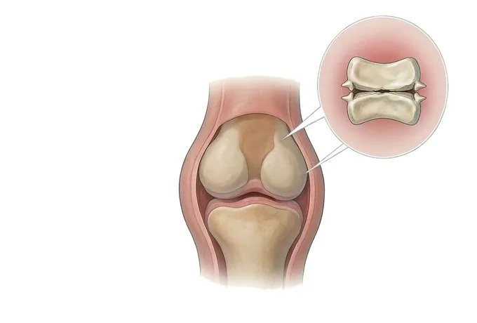
A joint is not a hinge with a lining of Teflon. It is a living organ with four working parts: the cartilage that lets two bone ends glide, the dense bone directly beneath it that absorbs what the cartilage passes on, the joint lining that produces a few millilitres of lubricating fluid, and the capsule and ligaments that hold it all under tension. Osteoarthritis is what happens when all four change together over years: the cartilage thins and frays, the bone beneath it hardens and grows spurs at the edges, the lining becomes irritable and swells, and the capsule tightens where it should not.
Because "wear and tear" is the phrase everyone reaches for, the condition is often heard as a verdict: the joint is used up, and nothing can be done until it is replaced. That reading is wrong in both halves. The degeneration is real — cartilage has very little capacity to rebuild itself — but the symptoms are not proportional to the wear, and the parts of treatment that work best (strength, load, weight, alignment) act on the joint's environment rather than on the cartilage surface itself. Many joints with heavy wear on film stay quiet for years; others with modest changes dominate a person's day.
The pain has a recognisable rhythm, and it is close to the opposite of the inflammatory arthritis it is often confused with. Osteoarthritic pain is worse with use: it builds through a day of walking and standing, it is worst at the end of the day, and it eases with rest. Morning stiffness is real but brief — usually gone within half an hour. Inflammatory arthritis behaves the other way round: long morning stiffness, pain at rest, and night pain that is present from the start.
One more distinction belongs at the beginning, because patients in this section often carry both. Osteoporosis — bone that has lost density and breaks under small loads — and osteoarthritis — a joint wearing out under normal loads — are different diseases of different tissue. Having one does not prevent the other, and treating one does not treat the other: a patient can need bone-protection medication and a knee replacement at the same time. For the bone-strength disease, see the separate guide to osteoporotic vertebral fracture.
Where it comes from: primary, secondary and by compartment
Osteoarthritis is classified first by whether the joint reached this state through ordinary load alone, and second — for the knee — by which compartment carries the wear. Both labels change what treatment is offered, which is why they are established before any decision about surgery.
| Type | How the joint arrived here | What it means for treatment |
|---|---|---|
| Primary | Ordinary load on a joint whose biology and shape offered no particular weakness, with age as the main driver | Progress is usually slow. Non-operative care has the widest window, and surgery is decided on function alone |
| Post-traumatic | An old injury — a meniscus removed, a ligament torn, a fracture that entered the joint — changed the joint's mechanics years ago | Wear can arrive a decade or more early and may be confined to one part of the knee, which opens options short of full replacement |
| Shape-driven | The joint was built differently — a shallow hip socket (dysplasia), a cam or pincer impingement, marked bow-leg or knock-knee | Alignment and shape can sometimes be corrected surgically before the joint is worn out, which is why shape is assessed early |
| Inflammatory-overlap | Years of low-grade inflammatory disease, or an old infection, damaged cartilage and left secondary degeneration behind | The inflammatory disease must be controlled first or alongside — replacing a joint while it is active invites failure |
For the knee, the compartment matters as much as the type. Most wear concentrates in the inner (medial) compartment, less often the outer, and sometimes only behind the kneecap. Wear confined to one compartment is the situation in which partial replacement and realignment osteotomy are discussed at all; wear across all three leaves total replacement as the anatomical option. The hip, being a ball-and-socket joint, is not divided this way — its surgical choice is between preserving the native joint (rarely, and early) and replacing it.
How severity is graded — and why the grade does not decide the operation
Two questions are asked together, and they are deliberately kept separate. The first is what the joint looks like: it is answered by the standing radiograph and described with the Kellgren–Lawrence grade. The second is what the joint does to the person: walking distance, stairs, sleep, work. Treatment decisions are driven by the second; the first explains it.
| KL grade | What the film shows | What it usually means |
|---|---|---|
| Grade 0 | A normal joint — full space, no bone reaction | Osteoarthritis is not the explanation for the pain; the search continues elsewhere |
| Grade 1 | Doubtful narrowing, small bone spurs at the edge | Early change. The moment to work on strength, weight and load habits — not a diagnosis of disability |
| Grade 2 | Definite spurs, clear but incomplete narrowing of the joint space | Established disease. Most patients here are managed non-operatively, and most can be |
| Grade 3 | Marked narrowing, hardening of the bone beneath the cartilage, spur formation | The zone in which function, not the film, decides between continued non-operative care and surgery |
| Grade 4 | Grossly narrowed or absent space, large spurs, deformity of the bone ends | Severe change — but even here, replacement is elected when the joint limits life, not because the grade says so |
The functional axis is what the appointment is actually about: how far can you walk, what do stairs cost you, is the pain present at rest or at night, has the knee begun to give way or the leg to look crooked. Two people with grade 3 films can sit at opposite ends of this axis — and it is the difference between a continued exercise programme and a scheduled operation. A grade that looks alarming on a film belonging to someone who walks five kilometres a day is not an indication; a joint that wakes its owner every night at grade 3 is.
How osteoarthritis is confirmed and what each test answers
The workup is short and deliberate: each test is chosen for the question it answers, and the order matters — the standing film comes first, and the more expensive studies are added only when the film leaves a question open.
| Test | The question it answers |
|---|---|
| Standing radiograph | Is the joint space narrowed, and how much? A film taken lying down hides most of the loss — weight-bearing is the standard, and both knees or both hips are filmed for comparison |
| Long-leg alignment film | Through which part of the knee does the weight pass? This is the study that makes osteotomy and partial-replacement decisions possible |
| MRI (selective) | Is something else or something more going on — a meniscus tear, early avascular necrosis, or symptoms far ahead of what the film shows? |
| Blood tests | Is this inflammatory arthritis after all? ESR, CRP and rheumatoid markers settle the question that the history usually already suggests |
The examination in between looks at what the radiograph cannot show: the way you walk, whether the knee bows when you stand on it, the range the joint still has, whether it holds against sideways stress, and where exactly the joint line is tender. Putting the film and the examination together produces the thing the decision actually needs: a statement of which structure explains which symptom, and what portion of the limitation is likely to yield to non-operative care.
Why patients travel to China for joint replacement
Joint replacement is one of the most standardised operations in surgery, and that is precisely why differences between providers matter: when the technique is mature, the things that separate outcomes are planning accuracy, surgical volume, anaesthesia and recovery management, and cost.
Three of those are the reason patients consider China. First, planning and alignment: our centres use robotic-assisted planning for knee replacement, which turns the implant position from an estimate made in theatre into a plan made before the skin is cut. Second, recovery management: enhanced-recovery pathways — regional anaesthesia where suitable, early standing the same day, structured rehabilitation from day one — are standard rather than exceptional. Third, cost: complete treatment in China typically runs at 30–50% of comparable care in Europe or the United States, with the implants, theatre standards and monitoring that the operation requires.
The practical side is arranged around that. Records reviewed by a joint-replacement surgeon receive a written opinion within about 48 hours of complete records arriving; we work with more than 30 commercial insurers for direct billing and have received patients from 124 countries and regions. Where the review concludes that an operation is not yet indicated — a common outcome at grades 1–2 — that conclusion is written down together with what should be tried first, and for how long.










