


Joint Stiffness & Cartilage Injury Overview
Joint Stiffness & Cartilage Injury Overview
Two problems share this page because they share a clock. Joint stiffness — most often the overgrowth of scar tissue inside or around a joint after surgery or injury — unlocks most easily while it is young: the window in which simple measures and one short anaesthetic work best is measured in weeks. A focal cartilage defect is the opposite in mechanism but the same in lesson: cartilage has no blood supply and cannot heal itself, and a defect can be repaired only while the joint around it is still healthy. Neither window closes suddenly, and neither closes without warning — this guide is about seeing them coming.
-
12 weeks
The window in which manipulation under anaesthesia works best for post-operative stiffness
-
48 hours
Written second-opinion report after complete records arrive
-
30+
International insurers working with us on direct billing
-
124+
Countries and regions our international patients come from
What stiffness and cartilage injury mean
Joint stiffness is a loss of the joint's range of motion. The commonest serious form after surgery or injury is arthrofibrosis: dense scar tissue growing inside and around the joint, binding the surfaces and filling the gutters through which the joint glides. It is worth saying what stiffness is not: it is not simply pain limiting motion, and it is not the normal slowness of a healing limb. It is a mechanical loss that a goniometer can measure — and it has a real cost, because a knee that will not fully straighten makes every step a negotiation, and a few degrees of lost extension change a gait more than thirty degrees of lost flexion do.
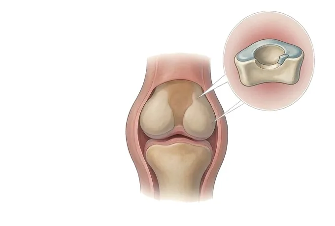
A focal cartilage defect is a hole in the bearing surface. Articular cartilage is the slick, load-bearing layer that lets bones glide against each other; it has no blood supply, and a full-thickness defect cannot heal itself the way skin does. Left alone, a focal defect tends to enlarge under load — the road that eventually leads to diffuse cartilage loss. That destination has a familiar name: osteoarthritis, and the distinction between focal and diffuse is the most important single fact in this field, because it decides whether the cartilage can be repaired at all.
The two problems meet in one place: both are easiest to treat early, and both announce themselves in ways a patient can notice — a joint that no longer straightens, a knee that catches or swells after activity it used to absorb. Neither is an emergency; both are appointments worth keeping.
Two problems, one clock: how the options close
A stiff joint is not one condition. The motion can be blocked by scar tissue (arthrofibrosis), by a tight capsule, by a mechanical block such as a loose body or a displaced fragment, by muscle guarding and pain alone, or by the joint simply being arthritic. Each has a different treatment, and telling them apart is the assessment's job — which is why arthrofibrosis is called a diagnosis of exclusion: the treatable impostors, above all infection after a joint replacement (revision surgery is the answer there), must be ruled out before scar tissue is blamed.
The clock differs by cause. Stiffness that follows surgery responds best to an escalating ladder in the first three months: rehabilitation with good pain control, then manipulation under anaesthesia, then arthroscopic release. Once stiffness is long-established, the same operations achieve less, because the scar has matured and the muscle has shortened.
Cartilage follows a parallel logic of candidacy. The repair ladder — marrow stimulation, cartilage transfer, cell-based implants — works for focal defects in a joint whose other surfaces, ligaments and meniscus are sound, and whose alignment can be made even. The same operations have little to offer diffuse wear, which is why the assessment measures not just the defect but everything around it.
| What blocks the motion | Where the answer lives |
|---|---|
| Young scar tissue inside the joint | The stiffness ladder: therapy, manipulation while the scar is young, release beyond that |
| A tight capsule after immobilisation | Graded stretching and load — motion prescribed with pain control that makes it possible |
| A mechanical block (loose body, flap, fragment) | Arthroscopy; the block is removed rather than stretched past |
| Muscle guarding and pain alone | Pain control and reassured movement — the joint moves further once it trusts the range |
| A component, after joint replacement | The revision guide; releasing scar will not move a misplaced part |
| Diffuse cartilage wear | Realignment or replacement; repair techniques are not offered to a diffuse pattern |
Grading: how much motion, how deep the defect
Both halves of this cluster carry well-established grading systems, and both grades drive the plan.
| Axis | How it is graded |
|---|---|
| Motion loss (stiffness) | International consensus grades knee stiffness by flexion: mild 90–100°, moderate 70–89°, severe below 70°. Extension loss is recorded separately and treated as its own problem — a lag of a few degrees already changes the gait. Numbers are compared over time, because the trend matters more than one reading. |
| Cartilage depth (ICRS grade) | Grade I softening and fibrillation; II a partial-thickness defect under half the cartilage's depth; III a deep defect of more than half the depth or reaching the bone underneath; IV full-thickness loss with exposed bone. Repair decisions live mostly in grades III–IV. |
| Cartilage area | Defect size in cm² shapes which repair fits: roughly under 2 cm² suits marrow stimulation; 1–4 cm² suits cartilage transfer; larger full-thickness defects suit cell-based repair; very large defects may need donor (allograft) tissue. Area is measured on MRI and confirmed at arthroscopy. |
| The joint around the defect | Alignment, ligament stability, meniscal status and the cartilage on the opposing surface — the factors that decide whether a repair can survive. Diffuse wear in the same joint changes the conversation to realignment or replacement. |
| Function and time | What the joint stops the patient doing, for how long, and how many weeks have passed since surgery or injury — the axis that decides how urgent the next step on the ladder is. |
How a stiff or painful joint is assessed
The assessment is deliberately simple and deliberately ordered. Motion is measured and written down as numbers, both flexion and extension, and compared with any earlier record. Weight-bearing radiographs show the joint space, alignment and any bone block. MRI with cartilage-sensitive sequences is the examination on which cartilage decisions rest: it shows the depth and area of a defect, the health of the surrounding cartilage, and the presence of scar bands in a stiff joint. Where stiffness follows a joint replacement, blood inflammatory markers and joint aspiration come before any diagnosis of scar tissue, because infection can present exactly this way, and CT checks the position of the components, because a component placed outside accepted angles can itself be the cause of stiffness (how a painful replacement is assessed carries that story in detail).
The output is written, and it answers three questions: how much motion is lost, what is blocking it, and whether the cartilage problem is focal and repairable or diffuse. Those three answers map directly onto the treatment ladder — and they are the difference between an operation chosen and an operation defaulted into.
Why patients travel to China for these operations
Both halves of this cluster reward systems more than single procedures. Our centres pair robotic-assisted planning for joint replacement and an orthopaedic 3D printing centre — established with the Shanghai Ninth People's Hospital team — with arthroscopic surgery and structured, staged rehabilitation built around nine standardised rehabilitation nodes. Assessment is centralised before treatment: motion measured as numbers, infection excluded before scar tissue is blamed, alignment measured before any cartilage repair is promised, and a written second opinion returned within about 48 hours of complete records arriving. Complete treatment in China typically runs at 30–50% of comparable care in Europe or the United States, with 30+ international insurers on direct billing.
The practical consequence for a travelling patient: the ladder is executed on schedule, because the schedule is the treatment. Rehabilitation, manipulation and any surgical stage are planned as one episode before the journey begins.










