


Pelvic & Acetabular Fracture Overview
Pelvic & Acetabular Fracture Overview
The pelvis is not one bone but a ring, and the socket of the hip sits in the side of that ring. A fracture of the ring and a fracture of the socket are two different injuries, and they are judged by two different questions. For the ring, the question is whether it still holds its shape and whether it is bleeding, which is why this injury is treated as a trauma emergency. For the socket, the question is whether the cartilage surface is still smooth and whether the weight-bearing roof still carries load, because a step left behind in that surface is what leads to arthritis later. Both usually follow a high-energy force in a younger person, and both can follow an ordinary fall in an older adult whose bone has thinned.
-
48 hours
Written opinion from our orthopaedic and trauma team once your images and records arrive complete
-
Two questions
Is the ring still stable, and is the joint surface still smooth — they are answered by different images
-
CT with 3D
The planning scan for both injuries: a plain film begins the assessment, it does not finish it
-
One plan
Trauma, reconstruction and rehabilitation reviewed together, including the late work if arthritis follows
What a pelvic fracture is, and why it is not the same injury as a broken socket
Two words get used interchangeably and should not be. The pelvis is a ring of bone: the sacrum at the back, and two hip bones at the sides and front, each made of three bones that fuse in childhood — the ilium at the top, the ischium below and behind, and the pubis at the front. The two hip bones meet each other at the front at the pubic symphysis, and each meets the sacrum at the back at a sacroiliac joint. Those joints are held by very strong ligaments, and together with the bones they form a closed ring whose job is to carry the weight of the upper body into the legs and to protect the bladder, the bowel and, in women, the reproductive organs.
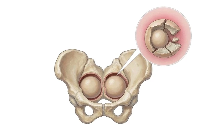
The acetabulum — the socket of the hip joint — is not a separate bone. It is a cup-shaped surface on the outer side of the hip bone, formed where the ilium, ischium and pubis meet. It is usefully thought of as two struts of bone arranged like an inverted Y: the anterior column in front (the iliopubic column) and the posterior column behind (the ilioischial column). The curved roof over the top of the cup is the weight-bearing dome, and it is the part that actually carries load when you stand and walk. The ball at the top of the thigh bone sits in that cup, covered in the same smooth cartilage.
So the two are injuries to different structures, judged by different questions. When the ring breaks, what matters is whether it still holds its shape — whether one break leaves it intact, or whether a second break or a torn joint lets part of it move — and whether it is bleeding, because the broken surfaces of spongy bone and the veins along the inside of the pelvis can lose a great deal of blood. When the socket breaks, bleeding is rarely the main problem; what matters is whether the cartilage surface is still smooth and the dome still carries load, because a step left in that surface is what wears the joint out over the years that follow.
That difference sets the tempo. An unstable, bleeding pelvic fracture is a trauma emergency: the pelvis is closed with a binder or a frame in the first minutes, bleeding is controlled, and the bone is dealt with once the patient is stable. An acetabular fracture is a scheduled operation planned from a CT scan, and it can wait a few days for swelling to settle. The phrase fracture of the pelvis on a discharge summary may describe an injury that needed nothing more than rest, or one that nearly killed the patient — the difference is not in the words but in the stability of the ring and the blood loss.
It also helps to know what these are not. A hip fracture is a break of the thigh bone below the ball, not of the pelvis or the socket. A ball that dies without a fracture is avascular necrosis of the femoral head, and a worn joint that was never injured is hip osteoarthritis.
The types: how the ring breaks, and how the socket breaks
Both injuries are named by the direction of the force that caused them, because the direction predicts what has torn and therefore what will move. The two schemes below are the ones used in practice.
| Type | What the force does | What it means in practice |
|---|---|---|
| Lateral compression (LC) | A blow to the side of the pelvis, or being run over from the side, pushes the ring inward and closes it | The commonest mechanism. The front of the ring breaks through the pubic rami and the back of the ring is crushed or the sacrum is fractured. The ring tends to shorten rather than open, and it is often stable vertically |
| Anteroposterior compression (APC) | A force from the front or back separates the two halves of the ring, classically described as an open book injury | The symphysis splits apart and the sacroiliac joints open. The whole ring is wider than it should be, which is the shape that bleeds. The greater the separation, the more the posterior ligaments have torn |
| Vertical shear (VS) | A vertical force, as in a fall from height landing on one leg, drives one half of the pelvis upward | The most unstable pattern. Half the pelvis with the leg attached is displaced upward, and the leg on that side may look short. These injuries are fixed rather than left to settle |
| Combined mechanism (CM) | More than one direction of force acting together | The pattern is mixed, and the plan is built from what the CT shows rather than from the label alone |
| Fragility fracture of the pelvis | An ordinary fall from standing height, in bone that has thinned | The commonest pelvic fracture in older adults. Usually the pubic rami, sometimes extending into the sacrum. It is painful and disabling but rarely bleeds dangerously; the problem is that it can progress and become a displaced fracture if a person keeps walking on it |
| Acetabular: elementary patterns | A single wall or a single column breaks — most often the posterior wall in a dashboard injury, but also the anterior wall, either column alone, or a transverse break across the cup | One component of the cup is separated. The posterior wall pattern is the commonest, and it is the one that travels with a dislocated hip |
| Acetabular: associated patterns | Two or more elementary patterns together — T-shaped, transverse with a posterior wall, a column with the opposite hemitransverse, or both columns | The cup is broken into several pieces and the joint surface is no longer one continuous curve. Both-column injuries separate the socket entirely from the rest of the hip bone while the joint surface itself stays roughly in position, which makes them deceptive on a plain film |
Two details matter more than the rest. An open book injury is the pattern to take most seriously in the first hour, because a ring that has opened has more room in it, and more room means more bleeding. And an acetabular fracture is very often accompanied by the ball of the thigh bone being driven out of the socket — a dislocation — which is itself an emergency, because the head of the bone loses its blood supply while it sits out of place. How the force reaches the bone is on the causes page, and the operations are on the treatment page.
How severity is read: stability of the ring, and smoothness of the joint surface
There is no single scale that covers both injuries, and trying to force them onto one is where most explanations go wrong. The ring is graded by mechanical stability; the socket is graded by how far the joint surface has moved and whether the dome is still doing its job.
| Grade | What it means | How it usually changes the plan |
|---|---|---|
| Tile A — the ring is stable | One bone is broken but the ring still holds its shape; the ligaments that hold the back of the ring are intact | The fracture itself does not need an operation. The work is pain relief, protected walking, and watching for a fracture that starts to shift |
| Tile B — rotationally unstable | The ring can twist but not slide upward; the back of the ring is partly injured but the vertical support is preserved | The ring is closed and held, often with a frame at the front or screws at the back, so that the shape is maintained while it heals |
| Tile C — unstable in both planes | The ring twists and also shifts upward; the back of the ring has lost its vertical support | This is fixed, at the front and at the back, once the patient is stable enough for the operation. The leg length and the sitting balance depend on getting the vertical position right |
| Acetabulum — undisplaced or minimally displaced, joint congruent | The crack runs through the socket but the surface is still smooth and the ball still sits centrally under the dome | Often treated without surgery, protected from full loading for a number of weeks, with films repeated to confirm the position has not changed |
| Acetabulum — displaced, dome intact | A piece of the joint surface has moved out of position, leaving a step, but the weight-bearing roof is still attached and usable | Operated on, with the aim of restoring a smooth surface. Anatomic reduction of the cartilage is the strongest single thing that shapes how the joint does years later |
| Acetabulum — the dome is involved | The weight-bearing roof itself is broken or is no longer sitting above the ball | The most demanding pattern. The roof has to be rebuilt, and where the surface has been badly damaged the longer-term plan may be a joint replacement rather than a repair |
There is one more axis underneath both of these: the patient. The same pattern on a scan means different things in a twenty-five-year-old cycling to work and an eighty-year-old who fell getting out of a chair — and not only because of the bone. In an older adult the strength of the bone holding an implant, the medicines being taken, the heart and lung reserve, and whether the person was living independently before the fall change the plan as much as the fracture does. That is why our service has a surgeon and a physician review an older patient together. The whole assessment is on the diagnosis page.
How the injury is confirmed, and the scan that plans the operation
The assessment runs on two tracks at once, and the order matters. In an unstable pelvic fracture the resuscitation comes first. A pelvic binder is placed around the hips at the level of the greater trochanters — not around the waist, which does nothing — to close the ring and reduce the space it has to bleed into. If the patient does not respond to that and to blood products, the bleeding is pursued directly: contrast CT to find a bleeding artery, catheter angiography to plug it, or surgical packing in front of the peritoneum to compress the venous and bone surfaces that are the usual source. A patient can die from the bleeding before anyone has had time to think about the bone, which is why this injury belongs in a unit that does all of that in one place.
The imaging then runs in a fixed order. A single front-to-back radiograph of the pelvis is taken early and shows the two things that change management immediately: a symphysis that has opened, and one half of the pelvis sitting higher than the other. Two more views follow — an inlet view, taken with the beam angled down, which shows whether the ring has rotated in or out, and an outlet view, angled up, which shows whether half the ring has shifted upward. When the socket is the question, two oblique views named after Judet are added: the iliac oblique, which shows the posterior column and the front wall, and the obturator oblique, which shows the anterior column and the back wall. A plain film alone cannot separate one column from the other, which is precisely why the socket is so often under-read from a single image.
CT with thin slices and three-dimensional reconstruction is the study that plans the operation, and in modern practice almost every patient with either injury has one. It shows what a radiograph cannot: the state of the sacroiliac joints at the back, the exact shape of the articular surface, any fragment lying loose inside the joint, and the route an implant would have to take. It is also the study on which a physical model can be printed for a complex reconstruction to be rehearsed before the patient is opened.
Three things are looked for alongside the bone, because each is easy to miss and each changes the plan. Blood at the opening of the urethra, or a prostate sitting high on examination, may mean the urinary passage is torn — and passing a catheter blindly can turn a partial tear into a complete one, so the passage is imaged first. An open fracture, with a wound communicating with the pelvis, means the bowel or the birth canal may be involved, with a high risk of deep infection. And a neurological examination of both legs is recorded before any reduction and repeated afterwards, because the sacral and sciatic nerves run directly against the back of the pelvis. The full sequence is on the diagnosis page.
Why patients come to China for pelvic and acetabular fracture care
The honest starting point is the same one we give for a fresh hip fracture: an acute, unstable, bleeding pelvic fracture is not a condition to travel with. It has to be stabilised where the patient is. Where our network is genuinely useful is in what comes next, and in the cases that have already become complicated — an acetabular fracture treated without surgery that has healed in a poor position, a socket that has worn into arthritis years later, a ring united with one half sitting high and the leg short, or chronic pain from a sacroiliac joint that never settled.
The reconstruction side
Pelvic and acetabular work is planning-heavy surgery, and the plan is the thing that decides the outcome. Our orthopaedic teams hold the full range of devices for this region — reconstruction plates and screws for the columns and walls, percutaneous screw systems for the posterior ring, and external frames for damage control — and fixation and joint replacement sit in the same service, so a socket that cannot be repaired is assessed by the team that can also replace it. Where the anatomy is unusual, the operation is rehearsed in advance on a physical model printed from the scan data, produced in a 3D printing centre run jointly with the academician Dai Kerong team of Shanghai Ninth People's Hospital, and our orthopaedic robot is available where a screw must be placed along a narrow corridor of bone.
The whole-patient side
A high-energy pelvic injury is rarely only a pelvic injury, and the bone is often not the thing that determines how the first month goes. Every patient is assessed for the injuries that travel with it — inside the abdomen and chest, the urinary passage, the nerves — and for the older adult whose fracture came from thin bone, the admission ends in a bone plan rather than a discharge note: the choice and timing of osteoporosis treatment, a falls assessment with specific changes written down, and the imaging intervals built into a follow-up schedule. Rehabilitation is structured and continues after discharge, with the later stages completed at home.
The practical side matters too. Records and images are reviewed by an orthopaedic surgeon working with the trauma and rehabilitation teams, and a written opinion is returned within about 48 hours of complete records arriving, setting out what the scan shows of the ring and the joint surface, and whether the fracture is better served by reconstruction or by a joint replacement. Our network runs 18 hospitals, works with more than 30 commercial insurers for direct billing where cover allows, and has received patients from 124 countries and regions.










