


Intra-articular Fracture Overview
Intra-articular Fracture Overview
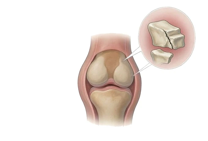
An intra-articular fracture is a break whose line runs into the cartilage surface of a joint — the knee, the ankle, the wrist, the elbow, the shoulder. It is not one disease but one pattern, and what binds these injuries together is not which bone is broken but one shared sequence: restore the joint surface so that no step remains, hold it with fixation strong enough to allow movement, and start moving before the joint stiffens. The difficulty at the centre of it is a contradiction, because cartilage survives on motion while bone unites on stillness. Two consequences follow that patients are usually not told. The measurement that matters is the step and the gap left in the joint surface, not the name of the fracture. And the day of the operation is usually set by the condition of the skin over the joint, not by the calendar.
-
What it is
A break whose line crosses the cartilage surface of a joint, in the knee, ankle, wrist, elbow or shoulder
-
The measurement that matters
The step and the gap left in the joint surface — not the name of the fracture pattern
-
The timing
The skin over the joint sets the day; open fractures, dislocations and threatened circulation are emergencies
-
The goal
A joint surface without a step, fixation that allows early movement, and rehabilitation started before the joint stiffens
What an intra-articular fracture is, and why the joint surface is the whole problem
Two bones meet at a joint, and the surfaces that meet are not bone. They are articular cartilage — a thin, glassy layer that has no blood vessels of its own and almost no ability to repair itself. It survives on the fluid inside the joint and on the movement that circulates that fluid. A fracture becomes an intra-articular fracture when its line runs into that layer. The bone underneath will heal; the cartilage it carried will not be replaced with anything like the original.
This is the reason the injury is judged by a different standard from an ordinary fracture. A break in the shaft of a bone can be accepted with the fragments a little out of line, because the bone will remodel and the limb will work. The same tolerance applied to a joint surface produces a step. Cartilage cannot redistribute load the way bone does: a step concentrates contact stress onto the edge of it, and the cartilage on that edge wears.
The second property is that cartilage needs movement. A joint held completely still loses range quickly: the capsule shortens, the surfaces adhere, and the cartilage thins through disuse. So the treatment has to satisfy two requirements that pull against each other — the surface must be restored accurately, which means operating on it, and the joint must be moving soon afterwards, which means the fixation has to be strong enough to be trusted while it moves.
It is worth separating this pattern from the fractures that sit next to it on the same skeleton. A fracture of the neck of the thigh bone is also a fracture near a joint, but the question there is the blood supply to the ball of the hip, and the answer decides whether the head is kept or replaced — the joint surface itself is usually intact. A fracture of the pelvis or of the hip socket is a problem of the pelvic ring and of the socket's anatomy, and it is assessed and treated as a different injury.
One more thing shapes everything that follows. The bone under the joint surface is often crushed rather than simply cracked, and the skin and muscle over it are injured at the same moment. The operation therefore has to be planned around two tissues that are both temporarily unfit: a joint surface that has lost its shape, and a soft-tissue envelope that has to recover before it can be cut through safely. That is why the plan is made from a CT scan, and why the operation is often deliberately delayed for days.
The joints it happens in, and the one boundary that matters
An intra-articular fracture is named after the joint it happens in, and the joint decides the exposure, the implant and the rehabilitation. What does not change between them is the standard: the articular surface is reduced, and the reduction is held.
| Joint | What is usually broken | What makes it difficult |
|---|---|---|
| Knee — tibial plateau | The flat top of the shin bone, often depressed downwards by the femoral condyle driven into it | A depressed segment rather than a displaced one: the surface has to be lifted back up and supported from beneath, and the meniscus and ligaments are assessed alongside |
| Knee — femoral condyles | The rounded lower end of the thigh bone, sometimes split between the two condyles | The fragments carry a curved surface that must be rebuilt as a curve, and the fracture often extends upwards into the shaft |
| Ankle | The malleoli on either side, the plafond above them, or all three | The joint is a tight mortise: a millimetre of displacement changes how the surfaces meet. The skin over it is thin, and the soft-tissue window is the narrowest of any joint |
| Wrist — distal radius | The lower end of the forearm bone where it meets the carpal bones | Common and easily underestimated: the joint surface can be involved in a fracture that looks unremarkable on a single view, and stiffness of the fingers is the complication to prevent |
| Elbow — distal humerus | The lower end of the upper arm bone, between the two condyles | A small joint with a very low tolerance for stiffness; the fragments are often thin and the bone in older patients fragile |
| Shoulder | The socket or the head of the humerus, or the greater tuberosity beside it | Stiffness dominates the recovery, and the choice between fixation and replacement depends on the blood supply to the head and on the patient's demands |
The boundary worth stating plainly is the hip. A fracture through the socket of the hip joint — the acetabulum — is an intra-articular fracture by this definition, but it is assessed as a fracture of the pelvic ring, with its own anatomy, its own classification and its own surgical approaches, and it belongs to the pelvic and acetabular page rather than to this one. The distinction is not pedantic: the socket is not exposed or fixed like a plateau or a malleolus, and the operations have almost nothing in common beyond the principle of restoring the surface.
Two further labels cut across the list. A fracture is open when the bone has communicated with the outside through a wound, which changes the timing from planned to immediate. And a fracture-dislocation is one where the joint has come out of its socket as well as broken, which makes the reduction an emergency in its own right. Both are dealt with under timing, because what they change is the clock.
How severity is read: the step, the pattern, and what the patient needs the joint to do
Three things are read together, and none of them alone is enough.
The first is the pattern. The AO/OTA classification sorts these fractures by how much of the joint is involved: type A is outside the joint, type B is partly articular — part of the surface is still attached to the shaft — and type C is completely articular, with the articular segment separated from the shaft entirely. The value of the letter is that it tells the surgeon how many separate articular fragments to expect and how much of the surface is free.
The second, and the one that matters more, is the step and the gap in the joint surface. Cartilage tolerates neither. A step leaves an edge that carries excess load; a gap leaves exposed bone and allows the surfaces to move abnormally against each other. This is the measurement the surgeon is actually trying to correct, and it is why the operation is described as restoring the surface rather than as fixing the bone. It is also the measurement that explains the apparent paradox of these injuries: a fracture that looks dramatic on a radiograph but leaves the surface flat may do better than a modest-looking one that leaves a step.
| What is read | What it decides |
|---|---|
| AO/OTA type A — extra-articular | The joint surface is intact. Standard fracture principles apply, and the joint itself is not the problem |
| AO/OTA type B — partial articular | Part of the surface remains attached and is used as the reference for reducing the rest |
| AO/OTA type C — complete articular | The surface is free of the shaft and has to be reconstructed from its fragments; the plan depends on the CT appearance |
| Step and gap in the articular surface | The single measurement most predictive of later arthritis. Reduction aims to leave neither |
| The soft-tissue envelope | Whether the joint can be operated on through the skin now, or whether the limb is stabilised and the operation deferred |
The third is the patient, and it changes the target rather than the technique. What the patient needs the joint to do afterwards sets how hard the reconstruction is pursued. a young manual worker who needs to kneel and lift requires a different standard from an older patient who walks on the flat. Age, bone quality, the condition of the joint before the injury, the demands of work and sport, and whether the injury is in a dominant limb all enter here.
Finally there is the time axis, and it runs the opposite way from the fracture most people have heard of. In a hip fracture the rule is to operate as soon as the patient is medically fit. In an intra-articular fracture of a limb, the joint is usually splinted or held with an external frame and the definitive operation is deliberately waited for — until the swelling has subsided and the skin wrinkles again, which is the sign that the soft tissues can be cut through and closed safely. Operating through blistered, tense skin is one of the few ways to turn a recoverable injury into a serious one. The waiting is part of the treatment.
How it is confirmed, and why CT is the image that plans the operation
The diagnosis is usually straightforward. A joint that is swollen, unwilling to move and painful after an injury is imaged, and radiographs taken in two planes at right angles show whether the fracture enters the joint. What the radiographs cannot do is show the operation.
That is what CT is for, and in this injury it is not an optional extra. A joint surface is three-dimensional: a fragment can be depressed rather than displaced, rotated rather than shifted, and hidden behind the fragments in front of it. CT with reconstructions in several planes shows how many articular pieces there are, which part of the surface is depressed and by how much, and which direction the fracture lines run.
MRI answers a different question. It shows cartilage, the menisci and the ligaments of the knee, the triangular fibrocartilage of the wrist, and the occult fracture that radiographs have not shown — because a fracture that has not displaced leaves no line for an X-ray to see and shows as bruising inside the bone. It is not the planning image for the bone, and it is not needed for every patient; it is used when the findings and the radiographs disagree, when a soft-tissue injury is suspected alongside, or when an undisplaced articular fracture is being looked for.
| Imaging | What it answers | What it does not do |
|---|---|---|
| Radiographs in two planes | Is there a fracture, does it enter the joint, and how displaced is it | It cannot show the depressed articular segment, the number of fragments or the rotation of a piece |
| CT with reconstructions | The articular fragments, the depression, the fracture lines — it plans the operation | It does not show the cartilage surface itself, the menisci or the ligaments |
| MRI | Cartilage, menisci, ligaments, and the undisplaced fracture that radiographs miss | It is not the planning image for the bone, and it is not required in every case |
Alongside the imaging, two examinations matter as much and are sometimes forgotten. The soft tissues over the joint are inspected at every review: the degree of swelling, the presence of blisters, whether the skin wrinkles again when the joint is moved, and whether there is a wound that communicates with the bone. And the circulation and nerves beyond the joint are checked before and after any reduction, because a fracture-dislocation can injure an artery or stretch a nerve, and a missed vascular injury costs more than any imaging finding.
What comes out of this is a plan with named parts, which the patient can ask about by name: what the articular fragments look like on CT, which approach will be used, whether the operation is immediate or staged, how the joint will be held in the interval, how much weight the limb may take afterwards, and when movement starts.
Why patients come to China for an intra-articular fracture
The honest starting point: an open fracture, a dislocated joint, a threatened limb or a suspected compartment syndrome is treated where the patient is, immediately, and not travelled with. What patients come for is everything that follows — and often the reconstruction of a joint that was operated on elsewhere and has not done well.
Planning and implants
These are the fractures where the operation is planned rather than improvised. The joint surface is reconstructed from a CT appearance, and the plan is drawn before the patient enters the theatre — the order of reduction, the approach, and the plates and screws needed to hold a surface that has been lifted back into place. Our network holds the full range of trauma and joint-reconstruction implants, including the anatomical plates and the fragment-specific hardware these patterns require, and three-dimensional planning is used where the pattern is complex or the joint surface is comminuted. Reduction is assisted with an arthroscope in selected patterns, so that the surface can be seen while it is reduced rather than judged from the radiograph afterwards.
Getting the joint moving
The part of the treatment that determines the eventual result happens after the operation. Our teams work to a written rehabilitation programme rather than a general instruction: how much movement and in which direction, how much weight the limb may take, and how both are advanced as the follow-up imaging allows. This is the part most often lost when a patient is discharged, and it is the part that decides whether a joint that was accurately reconstructed ends up moving. Where a joint has already stiffened after treatment elsewhere, or where a previous reduction has left a step, assessment and reconstruction are what patients travel for.
The practical side is arranged in the same way as for other conditions: the images are reviewed first and a written opinion is returned once the file is complete, with an estimate that states what is included and what is not. Direct billing is arranged with more than 30 commercial insurers where the plan and the authorisation allow it. Our network spans 19 institutions and treats patients from more than 124 countries and regions, and records reviews are returned in about 48 hours once the imaging and the history are complete.
One thing is worth saying about the second opinion, because it is what most of these enquiries turn out to be. A joint fracture that has healed with a step, or a joint that has stiffened during a long immobilisation, is often still treatable — the step can be corrected and the alignment restored, sometimes years later. The assessment starts from the same two images: a CT to see what the joint surface looks like now, and a weight-bearing radiograph to see how the joint is loading.










