


Hemifacial Spasm Overview
Hemifacial Spasm Overview
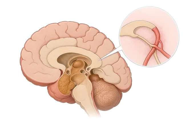
Hemifacial spasm is involuntary twitching of one side of the face. It is caused by irritation of the facial nerve, not by anything happening in the brain, and it has a shape that is the diagnosis: it begins around one eye , spreads downward over months to years to reach the cheek and the corner of the mouth, stays on one side , and continues during sleep . Those three features separate it from what it is routinely mistaken for. In most cases the cause is a blood vessel lying against the nerve where it leaves the brainstem, which is why the decisive scan is one that shows the nerve and the vessels rather than a routine one. This guide follows the order people need it in: what the condition is, the patterns it is confused with, the features that change the picture, how the assessment is assembled, and the two treatments — which are not two versions of the same thing.
-
48 hours
written opinion once complete records arrive
-
One team
neurology, neurosurgery and rehabilitation planning together
-
Pattern first
the diagnosis is made from the history and examination
-
124+
countries and regions our international patients come from
What hemifacial spasm is
Hemifacial spasm is involuntary, painless twitching of one side of the face. The cause is irritation of the facial nerve — the seventh cranial nerve, which supplies the muscles of facial expression — rather than anything happening in the brain itself. The muscle moves because the nerve supplying it is being stimulated, and the movement is the nerve's response.
It has a shape, and the shape is the diagnosis. It begins around one eye, usually as an intermittent twitching of the lower lid. Over months to years it spreads downward, to the cheek and then to the corner of the mouth, and in some people to the platysma in the neck. It stays on one side throughout. It continues during sleep, which is one of the features that separates it from a habit or a tic, and it cannot be suppressed for more than a moment. Those four features are the ones worth knowing, because all four can be observed at home before anyone is seen.
Two things are worth stating plainly at the start, because they are the questions people actually arrive with. It is not dangerous — it does not affect the brain, it does not threaten anything else, and it does not become another disease. And it does not resolve on its own. What it does is spread, slowly, in the pattern described above, and that spread is part of the condition rather than a sign of deterioration. What that spread looks like in practice is set out on the symptoms page.
This is also one of the most consistently mislabelled conditions in clinic, and the mislabelling has a cost. Because the spasm is reliably worse with fatigue, stress and speaking in front of others, the explanation offered for years is frequently stress. Stress does make it worse. What stress does not do is produce a one-sided twitch that starts around the eye, spreads downward over years, continues in sleep and cannot be suppressed. The cost of the wrong label is the years spent with a name that carries no treatment.
The pattern that identifies it, and what it is mistaken for
The table below sets out hemifacial spasm alongside the conditions it is most often confused with, and for each one the feature that separates it and what that distinction changes. It is not a list to work through alone — it is the map of what an assessment is sorting between.
| Pattern | What it looks like | The feature that identifies it | What the distinction changes |
|---|---|---|---|
| Hemifacial spasm | Twitching starting around one eye, spreading down over months to years to the cheek and the corner of the mouth | One side only, spread over time, continues in sleep, cannot be suppressed | Two treatment tracks: injection for the symptom, decompression for the cause |
| Blepharospasm | Involuntary closure of both eyes, sometimes forceful enough to keep them shut, worse with bright light | Both eyes, and it stays in the eyes rather than spreading down the face | A different disease with a different treatment, and without a surgical option of the same kind |
| Facial tic or habit spasm | A movement that relocates around the face, often beginning in childhood, that can be held back for a time | Suppressible, and it moves to a different part of the face rather than spreading downward in one line | Not a nerve compression. For most people the assessment ends here, with an explanation |
| Synkinesis after facial palsy | The eye closing when the mouth moves, or the mouth pulling when the eye is closed, in someone who had a facial palsy | A history of facial weakness first, and the movement is triggered by another movement rather than spontaneous | Managed differently. The key is the history of palsy, which is the question most often not asked |
| Focal motor seizure | Brief, stereotyped movements, occasionally with impaired awareness or a march up the face | The same sequence every time, lasting seconds, rather than a spasm that builds and eases | Investigated and treated on an epilepsy pathway, not on this one |
| Benign eyelid myokymia | A fine rippling of one eyelid that comes for days or weeks and then stops, familiar to most people | Self-limiting, with no spread to the cheek or the mouth, and it settles entirely | Needs reassurance rather than investigation, and it is not the same condition at all |
One row in that table deserves pointing out, because it is the one most often missed in both directions. Benign eyelid myokymia is the twitch nearly everyone has had: fine, rippling, confined to one lid, and gone within weeks. It is not hemifacial spasm. But a person who has had it repeatedly is the person most likely to be reassured again when the real thing starts, and the one feature that should end the reassurance is a twitch that has spread beyond the eyelid or that has not stopped. How the distinction is made is set out on the diagnosis page.
When the picture is not straightforward
Hemifacial spasm is benign, and saying so is not the same as saying nothing needs checking. What follows is the short list of features that do not fit, each of which changes what the assessment is looking for. None of them means something is wrong; each means a question has been raised that should be answered rather than absorbed into the working label.
| Feature | What it looks like | What it warrants |
|---|---|---|
| Onset in the lower face first | Twitching that begins around the mouth and travels upward, rather than starting at the eye | Reassessment of the diagnosis. The usual direction of spread is downward, and this is the exception |
| Weakness between episodes | A droop or asymmetry on that side that persists when the face is at rest and not spasming | Imaging and reassessment. Spasm does not leave a deficit behind between attacks |
| Reduced hearing, ringing or fullness in that ear | Hearing that has changed on the same side as the spasm | Imaging of the cerebellopontine angle. This points at a structural cause rather than a vessel alone |
| Onset before forty, or both sides involved | A spasm beginning in a child or young adult, or appearing on both sides of the face | A fuller assessment, including imaging, because both are outside the usual picture |
| Pain with the movement | Sharp, electric, brief pain in the face, in the same distribution as the movement | A different condition, discussed on the trigeminal neuralgia page |
| Other neurological symptoms with it | Double vision, slurred speech, weakness elsewhere, or a change in swallowing | Urgent assessment. These are not part of hemifacial spasm, and they move the timescale |
| A spasm that has changed character after years of stability | The pattern altering in distribution, frequency or the muscles involved after a long stable period | Reassessment rather than the same plan continued. The old label is not a reason to skip the new question |
The list is short and it is checkable without equipment, which is the point of putting it here rather than in a consultation. What should not happen is the reverse: a benign label applied once and never revisited, while a feature that does not fit sits in the notes unremarked. The features above are also the answer to the question most people are never given an answer to — what would make this worth coming back about. They are repeated on the symptoms page.
How the assessment is assembled
The assessment is clinical, and the history carries more weight here than any test. What imaging does is answer the questions the history and the examination have raised — and one of the commonest findings in a second opinion is that a scan was performed, reported as normal, and never actually showed the anatomy in question.
| Step | What it establishes | What it does not do |
|---|---|---|
| The description of onset and spread | Whether the pattern is the one that identifies hemifacial spasm — the single most informative part of the assessment, and it is available before any appointment | Not a substitute for examination. It decides what the examination is looking for |
| A video recorded during an episode | The distribution and the character of the movement, seen rather than described, which frequently settles the question on its own | Not a diagnostic test in itself. It is worth more than a paragraph written afterwards |
| Examination of facial movement | Which muscles are involved, whether the forehead is involved on that side, and whether there is weakness between episodes | Does not demonstrate the vessel. That is what imaging is for |
| The other cranial nerves, and hearing on that side | Whether anything else is involved, which is how the minority of structural causes are found | Not a screen in search of a finding. It is done because it is the part that changes the plan |
| Whether it continues in sleep | A feature that separates spasm from a habit or a tic, and it is usually reported by someone who has seen it | Not something a person can report about themselves, which is why the question is asked of a companion |
| 3.0T MRI with sequences for the nerve and vessels | Whether a vessel is lying against the facial nerve at the brainstem, and whether there is a structural cause behind it | Not every scan shows this. A study performed without the dedicated sequences can be normal while the anatomy was never displayed |
| Magnetic resonance angiography | Whether a vascular loop, an aneurysm or an unusual vessel is part of the picture | Not an addition to a normal scan with no indication. It answers one question, when that question is real |
| Electromyography | Whether the pattern is synkinesis rather than spasm, where the diagnosis is unclear or a palsy has preceded it | Not needed for a straightforward case. It is used where the two cannot be told apart clinically |
What the assessment ends in is a written judgement of four things: whether the pattern is hemifacial spasm or one of the four it is mistaken for; whether a vessel has been demonstrated and, if it has not, whether the scan performed was one that could have shown it; whether any feature of the atypical list is present; and what the first step is — which is sometimes an explanation and nothing more. The full assessment, including what it costs and how long it takes, is set out on the diagnosis page.
Why patients travel to China for a facial spasm assessment
Patients come for four reasons: a one-sided twitch that has been called stress for years and never given a name; an injection that has never been offered, or that was given once and produced a droop that nobody reviewed; a scan reported as normal that was never performed with the sequences that show the nerve and the vessels; or a question about whether decompression is now the right step rather than another decade of injections. The value here is not a different machine. It is a team that runs neurology, neurosurgery and rehabilitation in one pathway, and that says which pattern this is and what has never actually been looked at.
What the pathway looks like. Neurology, neurosurgery and rehabilitation plan together. 3.0T MRI, dual-source CT, PET-CT, 3D DSA and long-term video EEG sit inside the same programme, as do electromyography and formal hearing assessment. A written opinion is returned within 48 hours of complete records arriving. Fully treated costs typically run at 30–50% of comparable care in Europe or the United States, more than 30 commercial insurers are billed directly, and our international patients come from 124 countries and regions.
What must be said about the operation. Microvascular decompression is a specific operation with a specific place in this pathway: it is considered where the spasm is costing a person enough in their working life, where a vessel has been demonstrated, and where the person accepts what an operation involves. It requires general anaesthesia and a hospital stay, and it carries the risks that any posterior fossa operation carries, including effects on hearing on that side. We name it with its place in the pathway rather than as a promise, and anything recommended that is not carried out here is named at the outset rather than at the end of a workup, before any payment is discussed. For a large proportion of people, the injection is doing enough, and that is said as plainly as the alternative.
What a records-only opinion can and cannot do. It can say whether a description fits hemifacial spasm or one of the patterns it is mistaken for, whether the imaging that was done was the imaging that shows the relevant anatomy, whether an atypical feature is present, and whether either treatment is worth discussing. It cannot examine the person, and examination is where the diagnosis is made — the movement itself, the muscles involved, and whether there is weakness between episodes all require a person in the room. We say so rather than implying otherwise.
What to send first. A short note of which eye it started in, how long ago, and how far it has spread; a phone video recorded during an episode, which is frequently the most useful single item in the file; whether anyone has seen it during sleep; and any previous scans together with their reports, so that it is known which sequences were performed. That is the part of the file no hospital can reconstruct later.










