


Hallux Valgus & Foot Deformity Overview
Hallux Valgus & Foot Deformity Overview
This page is about feet that have changed shape, and about the three questions that decide what is done about them. The first is whether the alignment is actually changing over time or has been stable for years, because a deformity that has not moved does not need an operation merely because it is visible. The second is where the pain is coming from: the size of a prominence and the severity of the pain are only loosely related, and the pain may sit in the joint, in the skin over the bump, or under a different part of the foot. The third is whether the deformity is flexible, meaning it can still be brought back towards a normal position by hand, or fixed, meaning it cannot — which decides whether a shoe, a pad or an insole has any chance of helping. Hallux valgus and the wider family of foot deformities share these principles, and the specifics are set out on the pages that follow.
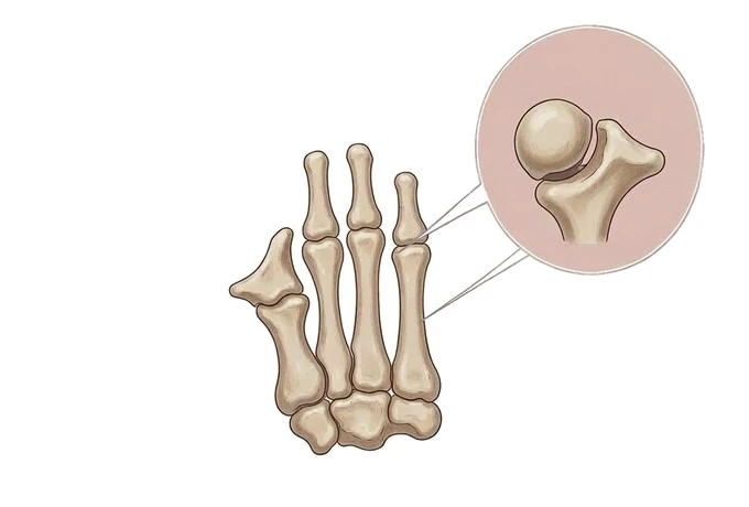
-
The bump is the edge of a joint, not a growth of bone
removing the prominence alone does not change the alignment that produced it
-
Pain and deformity are two separate questions
the size of the prominence and the severity of the pain are only loosely related
-
Flexible or fixed decides whether support can help
a foot that can be brought back by hand is managed differently from one that cannot
-
The great toe is usually not the only toe involved
load transferred away from the first ray is what makes the second toe curl
What hallux valgus and foot deformity are, and what the bump actually is
The word most patients use is “bunion”, and it describes a bump. The condition is not a bump: it is a change in the alignment of the whole first ray, and that distinction is the difference between a plan that works and a series of measures applied to the wrong thing.
- In hallux valgus the great toe drifts towards the second toe, and the first metatarsal moves away from it. The two movements happen together, which is why the joint at the base of the great toe sits proud on the inner edge of the foot, with the skin, the soft tissues and the small bursa stretched over it.
- The deformity is structural, and that word has a specific consequence. A shoe can be made wider, a pad can sit over a prominence, an insole can move load away from a sore spot, and each makes a foot more comfortable. None of them puts the joint back into line: materials change how a foot is loaded, not where the bones sit.
- Pain and deformity are two different measurements, and they do not track each other. A foot with a marked deformity may be entirely comfortable, and one with a modest deformity may be disabling. The pain may come from the skin and bursa over the prominence, from the joint as it takes an abnormal load, from the nerve stretched across it, or from elsewhere in the foot where load has been transferred. Finding which is responsible is why an operation is planned from the examination and the standing radiograph rather than from how the foot looks.
- Foot deformity is a family rather than a single condition, and the name decides the treatment. A curled lesser toe, an arch that has flattened in stance, an abnormally high arch and pain under the ball of the foot are all deformities, with different causes and different corrections. The first question after the examination is therefore which deformity is primary, because a second toe treated on its own while the first ray is left as it is tends to come back.
Why this matters when choosing where to be treated. The commonest experience of someone with a painful bunion is a long series of shoes, pads and insoles, each helping for a while and none changing the shape of the foot. Families who understand that alignment, pain and flexibility are three separate questions ask the ones that move a case forward: has the alignment actually changed since the last radiograph, which structure is producing the pain, and what exactly is going to be realigned if an operation is considered.
The kinds of foot deformity, and why the name comes before the treatment
Deformities of the forefoot are grouped by what has gone wrong, and the grouping decides which operation, if any, addresses the problem — including whether the standard bunion operation is the wrong one.
| Type | What is driving it | What this means for treatment |
|---|---|---|
| Hallux valgus, flexible | The first ray has lost alignment but the joint still moves and the toe can be brought back towards the midline by hand | Footwear and padding are the first measures; when an operation is needed it is directed at the part of the ray that has moved |
| Hallux valgus with an unstable first joint | The base of the first metatarsal moves abnormally, so the whole ray shifts rather than just the toe | An operation on the toe alone is likely to fail; the correction has to include the joint at the base of the metatarsal |
| Hallux rigidus | The joint at the base of the great toe has worn and become stiff, so it no longer bends upwards | This is a worn joint rather than a deviated one, and the operations that suit a deviated joint do not suit this |
| Lesser toe deformity | A hammer toe, a claw toe or a mallet toe, usually because the toe has taken load the first ray no longer carries | Correcting the toe without addressing the load transferred to it is why these deformities recur |
| Flexible flatfoot | The arch disappears when standing and returns when the foot is lifted, so the deformity is in the soft tissues | Support changes how the foot is loaded and can relieve symptoms; it does not create an arch |
| Rigid flatfoot and cavus | The shape is fixed, either from a joint that has stiffened or from an underlying neurological or structural cause | A fixed deformity is investigated for its cause before it is treated, and cannot be corrected by support |
| Metatarsalgia | Pain under the ball of the foot, often where load has been transferred from the first ray or where a metatarsal is long | Treated by redistributing load first; where the cause is a metatarsal, the treatment is directed at that bone |
| Inflammatory forefoot deformity | Inflammation attacking the joints of the forefoot directly, which changes the shape of the whole front of the foot | Controlling the joint disease is part of the treatment, and the operation chosen is adapted accordingly |
Two of these are worth separating in the mind, because they are the ones most often confused. A bunion and a stiff great toe are often described by patients in the same words and are not the same problem: one is a loss of alignment in a joint that still moves, the other a loss of the joint surface in a joint that no longer does.
Two axes: how far the alignment has moved, and what it costs the patient
Severity here is read on two axes at once, and they are not substitutes for each other. The first is anatomical: how far the alignment has moved, measured on a standing radiograph. The second is functional: what the foot can no longer do, which is what the patient experiences and what treatment is aimed at.
Axis one — the angles, measured standing. Two angles are measured from the weight-bearing film. The hallux valgus angle is the angle between the first metatarsal and the great toe, and the intermetatarsal angle is the angle between the first and second metatarsals. The values below are conventional groupings from the literature: they describe the deformity, not the patient, and are one input among several.
| Grouping | Hallux valgus angle | Intermetatarsal angle | What it usually means |
|---|---|---|---|
| Normal alignment | Under 15° | Under 9° | No deformity by measurement, whatever the shape of the foot suggests |
| Mild | 15–20° | Under 11° | A small movement of the toe; symptoms usually come from footwear rather than from the joint |
| Moderate | 20–40° | 11–16° | The deformity is established; the joint may be taking an abnormal load and the second toe may be involved |
| Severe | Over 40° | Over 16° | The whole ray has moved; the second toe is commonly affected and the joint may be losing its surface |
Three further measurements from the same film change the plan more often than the angles themselves: whether the joint at the base of the great toe is congruent (the surfaces still face each other) or incongruent (they have rotated apart); where the small sesamoid bones under the first metatarsal head sit; and whether the joint surface shows the narrowing and irregularity of wear. To these is added a finding no radiograph shows: how much the first ray moves at its base.
Axis two — what the foot can no longer do, recorded in the patient’s own terms. This axis decides whether treatment is needed at all.
- Where the pain is, and what brings it on. Pain from the upper of a shoe, relieved by taking the shoe off, points at the prominence and its bursa. Pain felt deep in the joint on walking or pushing off points at the joint. Pain under the ball of the foot or in the second toe points at transferred load. These are different problems, and they are not solved by the same measure.
- What the foot can no longer tolerate. The distance that can be walked, the time that can be spent standing, whether ordinary shoes can be worn for a working day, and whether activities that matter to the person have been given up. This is the most useful single record in the notes, because it can be compared after treatment.
- How the alignment has behaved over time. A deformity that has been the same for a decade is a different proposition from one that has moved over the past two years, and the comparison is made against earlier radiographs or photographs rather than against memory.
- What is happening to the toes beside it. A second toe that has begun to curl, a corn on top of a joint, or callus that has moved under the ball of the foot all indicate that the load pattern of the whole forefoot has changed, and they are often the reason the patient seeks help.
Reading the two axes together is what produces a plan. A severe angle with no pain and no limitation is watched; a mild angle with pain produced entirely by footwear is managed by changing the footwear. A moderate angle with joint pain, a second toe beginning to deform and a first ray that moves abnormally is the situation in which the choice of operation, and the part of the ray it is aimed at, becomes the central question.
What has to be established before anything is chosen
The assessment of a foot deformity is short and specific, and all of it can be done in a single visit. What it produces is a set of answers rather than a diagnosis, because the diagnosis is usually obvious from looking: the questions are how far, where from, and how fixed.
- How the foot behaves under load. The examination is performed standing as well as sitting. The alignment of the great toe, the position of the arch and the site of any callus are different in the two positions, and the standing position is the one that produces the symptoms.
- Whether the deformity is flexible or fixed. The great toe is brought back towards the midline by hand, and the amount of correction that can be obtained is recorded. A deformity that comes back easily is a deformity of soft tissue balance and can be affected by how the foot is loaded; one that cannot be moved has a fixed component that no shoe will change.
- Whether the first ray is stable at its base. The base of the first metatarsal is assessed for abnormal movement. This single finding is one of the strongest determinants of which operation will hold, and it cannot be seen on a radiograph.
- Whether the joint at the base of the great toe has lost its surface. Stiffness, a reduced range of upward bending and crepitus point to a worn joint rather than a deviated one, which changes the operation.
- What the patient actually wears. The shoes are examined and their width compared with the width of the standing foot. A mismatch between the two is often the whole explanation for the pain, and it is the one finding that can be acted on the same day.
- The load elsewhere in the foot. The lesser toes, the ball of the foot, the arch and the ankle are all examined, because a deformity of the first ray changes how the rest of the foot is loaded — and that is often where the patient feels it.
- Whether something other than mechanics is involved. Evidence of inflammatory joint disease, gout, a nerve entrapment between the metatarsals, or loss of sensation from diabetes changes the plan before any operation is considered, and each is looked for deliberately rather than assumed absent.
The one investigation that matters. The radiograph is taken with the patient standing and bearing weight on both feet. A foot imaged lying on a table is not the foot that hurts, because the deformity is a change in alignment under load, and non-weight-bearing images can understate it or miss it. From the standing film the angles are measured, the shape of the joint surfaces is recorded and the position of the sesamoid bones is noted. Cross-sectional imaging is not routine and answers a specific question: an MRI where a soft tissue or inflammatory cause is suspected, a CT where a complex deformity or a previous operation has to be planned in three dimensions.
What the assessment should end with. A statement of which deformity is primary; the measured alignment; whether the deformity is flexible or fixed and whether the first ray is stable; which structure produces the pain; and what the options are, including doing nothing. Where an operation is discussed, the record should say which part of the ray will be realigned and why that part.
Why patients travel to China for foot and ankle surgery
Forefoot surgery rewards planning more than any single technique, and planning is what a system can organise around the patient. Our orthopaedic services pair assessment built on weight-bearing radiographic measurement with an orthopaedic 3D printing centre — established with the Shanghai Ninth People’s Hospital team — and with arthroscopic surgery and structured rehabilitation built around nine standardised rehabilitation nodes. The alignment is measured before an operation is proposed, the part of the ray responsible is identified before the procedure is chosen, and a written second opinion is returned within about 48 hours of complete records arriving. Complete treatment in China typically runs at 30–50% of comparable care in Europe or the United States, with 30+ international insurers on direct billing.
The practical consequence for a travelling patient is that the plan can be arranged as one episode: the assessment, the choice of procedure, the operation and the protected weight-bearing period that follows are scheduled together before the journey begins. Where the deformity is on both feet, the timing of the two sides is decided in the same conversation, because the recovery of one foot constrains what can be done about the other.
Next in this guide: for how the alignment is measured and what the assessment has to answer, go to diagnosis: weight-bearing angles and the three questions; for what the treatment itself involves, read treatment: shoes, timing and the right operation.










