


Avascular Necrosis of the Femoral Head Overview
Avascular Necrosis of the Femoral Head Overview
Avascular necrosis is not arthritis. Arthritis is a joint wearing out under load over decades; avascular necrosis is bone dying because the blood that supplies it has been interrupted, and it can take a hip from normal to collapsed in months. It arrives most often between the thirties and the fifties, it very often involves both hips, and the difference between a preserved hip and a replaced one is usually decided by how early the diagnosis is made. This guide follows one idea throughout: before the bone collapses there is a window, and after it there is not.
-
48 hours
Written second-opinion report after complete records arrive
-
Both hips
Imaged on MRI in every case with a systemic cause
-
30+
International insurers working with us on direct billing
-
124+
Countries and regions our international patients come from
What avascular necrosis is — bone death, not arthritis
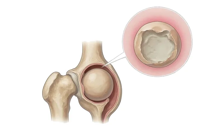
Bone is living tissue, and like every living tissue it needs blood. The ball of the hip — the femoral head — is supplied by a small number of vessels that run along the neck of the femur, and it has almost no second supply to fall back on. If those vessels are interrupted, by a fracture, by a clot, by pressure inside the bone, or by damage from steroids or alcohol, the bone in the head begins to die. That is avascular necrosis: literally, death of bone from loss of its blood supply. It is also called osteonecrosis, and older texts call it aseptic necrosis.
What happens next is mechanical rather than inflammatory. Dead bone keeps its shape for a while but loses its strength, so the thin plate of bone directly under the joint surface — the subchondral bone — starts to fatigue. A small crack appears under the surface, seen on a radiograph as a thin crescent of darkness. Once that crack gives way, the surface collapses, the ball is no longer round, and the joint that was normal months earlier becomes arthritic. This is why the disease behaves so differently from osteoarthritis: the cartilage is not the first casualty and the process is not driven by load. It is driven by blood supply, and it moves in months rather than decades.
Two consequences follow from this, and they organise everything else in this guide. The first is that the diagnosis is a race. In the earliest stage nothing is visible on a plain film, and a patient whose hip is examined and X-rayed but not scanned may be told there is nothing wrong until the head collapses — at which point the joint-preserving options have already gone. MRI is what stops that clock, and it is the reason the diagnostic threshold for a painful hip in a person with a risk factor is low.
One distinction belongs here, because it changes the whole conversation: avascular necrosis is not infection, and it is not a tumour. Both can destroy a hip and both can look similar on a single film, which is why the imaging sequence and, occasionally, a biopsy matter. And it is not the same as the wear-and-tear disease in the sibling guide to hip and knee osteoarthritis: a patient can have both, but only one of them has a window measured in months.
The second conclusion is that both hips belong to the same problem. When the cause is systemic — steroid treatment, alcohol, sickle cell disease, a clotting disorder — the other hip is involved in most patients, sometimes at the same time and sometimes a year later. A plan that deals with one side and does not look at the other is not a plan. This is why our imaging protocol always includes both hips, and why we ask patients to tell us early if the other side develops new pain.
Where it comes from, and where else it appears
Avascular necrosis is classified by what interrupted the blood supply, because that determines whether anything can be changed, and by how much bone is involved, because that determines how long a joint-preserving operation can be expected to hold. The four groups below cover almost every patient.
| Group | What interrupted the supply | What it means for treatment |
|---|---|---|
| Corticosteroid-associated | Steroid treatment — high-dose short courses as well as long-term low-dose treatment, and including steroid pulses given for other conditions | The medication is usually still needed, so it is reviewed rather than simply stopped; both hips are scanned; the window is often shorter than in other groups |
| Alcohol-associated | Sustained heavy drinking, which affects fat metabolism, blood flow and the cells that maintain bone | Reducing intake is the one lever that is fully in the patient's hands; it is also associated with more extensive and more often bilateral disease |
| Post-traumatic | A fractured neck of femur, a hip dislocation, or earlier surgery on the hip that damaged the vessels along the neck | The timing is predictable after the injury, so the hip is followed deliberately rather than left to symptoms |
| Secondary and idiopathic | Sickle cell disease, clotting disorders, lupus and other autoimmune disease, Gaucher disease, radiotherapy, chemotherapy, decompression sickness, HIV — or no cause found at all | Finding the cause changes surveillance and prevention; when nothing is found, the disease is still treated on its stage |
The second classification is anatomical. The femoral head is the site in the great majority of cases, which is why this guide is about the hip; but the same process occurs at the knee (most often the inner side of the femoral condyle), the shoulder (the humeral head), the ankle (the talus) and the small bones of the wrist, and those sites are usually associated with the steroid, alcohol and blood-related causes rather than with trauma. When more than one site or more than one joint is involved, the term is multifocal osteonecrosis, and it is a signal to look harder for a systemic cause and to image the other joints before symptoms appear.
For the hip there is one further description that matters to the surgeon but barely appears in the patient's experience: the size and position of the lesion within the femoral head. A small lesion on the inner side, carrying no load, behaves quite differently from one occupying the outer upper part, which is exactly where body weight passes through. Two femoral heads can carry the same stage number and face entirely different prospects — which is why the stage has to be read together with the measurement of extent.
Staging: ARCO, Ficat, and why collapse is the dividing line
Two questions are asked together and kept separate. The first is structural: has the joint surface collapsed yet? It is answered by the radiograph and by MRI, and it is described with the ARCO stage or the older Ficat and Arlet stage. The second is functional: what does the hip still allow, and how much pain does it cost? The first decides which operations are even candidates; the second decides when to move.
| Stage | What the imaging shows | What it means |
|---|---|---|
| ARCO 0 / Ficat 0 | All imaging normal in a hip with symptoms, or in a patient known to be at high risk or with proven disease on the other side | The disease is suspected rather than seen; MRI is repeated rather than the patient reassured and discharged |
| ARCO 1 / Ficat I | MRI abnormal — the typical low-signal line on T1 and the double-line sign on T2 — while the radiograph is still normal | The stage in which nothing is yet visible on a plain film. This is the stage the whole diagnostic strategy is designed to reach |
| ARCO 2 / Ficat II | Sclerosis and small cysts on the radiograph, no crescent and no flattening; the head is still spherical | Pre-collapse. Joint-preserving surgery is on the table, and the size and position of the lesion decide how much it can be expected to achieve |
| ARCO 3 / Ficat III | A crescent sign — a thin subchondral crack — with or without flattening. ARCO divides this into 3A (depression under 2 mm) and 3B (2 mm or more) | The turning point. Small depressions are still discussed for preservation in selected patients; once the surface has flattened, replacement is the reliable answer |
| ARCO 4 / Ficat IV | Collapse with joint-space narrowing, sclerosis of the socket and secondary arthritis of the whole joint | The joint itself is now the problem, not only the segment of dead bone. Replacement is the durable treatment |
Three things are worth saying about this table before any treatment is discussed, because patients read it as a verdict and it is not one. First, stage and pain are not the same thing: a small lesion can cause severe pain while a larger one, further from the weight-bearing surface, can be relatively quiet, and the reverse also happens. Second, stage alone does not choose the operation: a stage 2 lesion covering most of the weight-bearing surface has a worse natural history than a small stage 3A lesion in the same place, which is why the extent measurement sits beside the stage number in the report. Third, staging describes the hip, not the patient: the same stage in a person of thirty-five and in a person of sixty-five, or in a person with kidney disease who may need steroids again, leads to different advice.
The functional axis is the half the patient brings to the clinic: how far the hip can still walk before the pain stops it, how many hours of the day it hurts, whether it wakes them, how much passive internal rotation has been lost, and how fast all of this is changing. A hip that has gone from normal to needing a stick in three months, and a hip that has been stable at the same stage for two years, are two different clinical situations — and treatment is timed by this axis rather than by the number alone.
How the diagnosis is made, and what each test answers
Each test answers a different question, and the order matters: the wrong order costs the window.
| Test | The question it answers | What it cannot do |
|---|---|---|
| MRI | Is the bone dead, and how large is the segment? It shows the disease when the radiograph is still normal, and it grades the lesion | It is not a substitute for staging collapse; the thin subchondral crack and the depth of depression are read on plain films and CT |
| Radiograph of the pelvis and hip | The stage: sclerosis and cysts, the crescent sign, flattening, joint-space narrowing | It is normal in the earliest stage — a normal film does not exclude the diagnosis in a symptomatic hip |
| MRI of the opposite hip | Is the other side already involved, months before it hurts? | — (this is the examination that is most often skipped, and the one that most often changes the plan) |
| CT | The depth of any depression, and the bone stock available when a difficult replacement is planned | It adds little in a pre-collapse hip that MRI has already characterised |
| Blood tests and history | Whether there is a cause that can be treated or watched: lipids, clotting studies, haemoglobinopathy, autoimmune markers, steroid and alcohol history | They do not make the diagnosis; a normal set of tests does not mean the disease is idiopathic in origin |
Two points of practice follow. The first is that the sequence for a painful hip in a person with a risk factor is MRI first, and both hips at once — not a radiograph and a review in six months. The second is that the diagnosis is not complete until the report answers four things: whether the bone is dead, at what stage, how large and where the segment sits, and what the other hip is doing. A report that says only "avascular necrosis, right hip" leaves every treatment decision unmade.
Why patients travel to China for hip surgery
Hip preservation is a decision made from images and from time, and both are cheap to move. The imaging protocol is the same everywhere, the staging systems are international, and the two operations that matter — core decompression for the pre-collapse hip and total hip replacement for the collapsed one — are standardised procedures. What separates providers is how quickly a decision is reached, how carefully the pre-collapse hip is selected for preservation, and how the recovery is managed.
Records reviewed by a joint specialist receive a written opinion within about 48 hours of complete records arriving, and that opinion states the stage, whether preservation is still worth attempting, and which of the available operations applies. Where the review concludes that a hip is not yet at the point of needing surgery, that conclusion is written down together with what should be monitored and how often. For replacement, our centres use robotic-assisted planning for knee and hip replacement, which turns the position of the implant into a plan made before the skin is cut rather than an estimate made in theatre, and 3D-printed planning models — a capability developed with the orthopaedic 3D printing centre established with the Shanghai Ninth People's Hospital team — are used for complex and revision cases where the bone has already been distorted by collapse or by previous surgery.
Recovery is managed on an enhanced-recovery pathway: regional anaesthesia where suitable, standing and walking on the day of surgery, and structured rehabilitation from the first day, with nine standardised rehabilitation stages and discharge against a milestone list rather than a calendar date. Complete treatment in China typically runs at 30–50% of comparable care in Europe or the United States, with the implants, theatre standards and monitoring the operation requires. We work with more than 30 commercial insurers for direct billing and have received patients from 124 countries and regions.










