


Rotator Cuff Tear Overview
Rotator Cuff Tear Overview
A rotator cuff tear is one of the most common findings in the adult shoulder, and one of the most commonly misread. Imaging studies of people with no shoulder complaints find full-thickness tears in a substantial minority from the mid-fifties onward: the tear is real, but for many it never becomes the reason they seek care. The decision that matters is therefore not whether a tear is present. It is how large the tear is, how far the tendon has retracted from the bone it should attach to, and how much of the muscle has been replaced by fat. Those three measurements decide whether a repair is likely to hold, whether reconstruction is the more honest operation, or whether the cuff has moved past repair altogether. This guide is about reading those three numbers, and about the one situation in which waiting has a price.
-
65 years
Of continuous orthopaedic practice behind the centre treating these shoulders
-
48 hours
Written second-opinion report after complete records arrive
-
30+
International insurers working with us on direct billing
-
124+
Countries and regions our international patients come from
What the rotator cuff is, and what a tear means
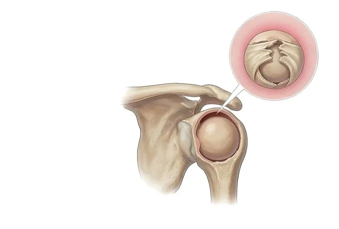
The rotator cuff is not a single structure but a sleeve of four tendons — supraspinatus, infraspinatus, teres minor and subscapularis — that wrap around the head of the humerus like a cuff around a shirtsleeve. Together they do two jobs: they centre the ball in its shallow socket while the arm moves, and they rotate the arm outward, inward and upward. The most commonly torn of the four is the supraspinatus, the tendon that passes through the narrow space beneath the acromion and lifts the arm away from the body.
A tear is a break in that tendon — partial, cutting into part of its thickness, or full-thickness, running right through. What surprises most patients is that the finding is so common. Imaging studies of adults with no shoulder pain find full-thickness tears in a large minority as age advances, and cadaveric series find them still more often. This is the first fact worth holding on to: a tear on a scan is not the same as a shoulder that needs an operation. Many tears are silent for years, and a good number never cause enough trouble to justify surgery.
The second fact is that not all tears arrive the same way. A tear that accumulates with age — the ordinary degenerative tear — has a long, forgiving history and can often be managed with rehabilitation and time. A tear caused by a fall onto the shoulder, or by a sudden pull on an outstretched or lifted arm, is a different problem: the tendon retracts from its attachment, the muscle begins to change, and the window in which a straightforward repair is possible starts to close. The two stories share a diagnosis and share almost nothing else.
Partial, full-thickness, and the three numbers that decide repair
The first distinction is depth. A partial-thickness tear involves part of the tendon's substance — at the joint surface, at the bursal surface, or within the tendon — and a particular pattern on the joint side of the supraspinatus is well described. A full-thickness tear runs through the whole tendon, so that the joint and the bursa above it communicate. Depth matters because the two behave differently: partial tears frequently improve with rehabilitation, while a full-thickness tear has no capacity to heal itself, because the tendon's blood supply at that point is already marginal.
Size is the second distinction, and it is the one most often quoted. Reported in centimetres of anteroposterior length, tears are conventionally grouped as small (under 1 cm), medium (1–3 cm), large (3–5 cm) and massive (over 5 cm). Size matters for a practical reason: the larger the tear, the greater the load each stitch must carry, and the more likely it is that published series will report the repair failing to hold.
But size alone decides nothing, which is why the two numbers that follow are the ones worth travelling with. Retraction describes how far the torn tendon end has pulled away from the bone — graded as sitting near its footprint, at the level of the humeral head, or down at the level of the glenoid. A 3 cm tear that has barely moved is repairable with far more confidence than a 3 cm tear whose end sits far medial. And muscle quality describes how much of the muscle belly has been replaced by fat, graded from 0 to 4. As fatty infiltration advances, the muscle can no longer generate force even if the tendon is stitched back, which is precisely why the same operation performed two years later achieves less than it would have earlier.
Read together, these three numbers describe a joint on a spectrum rather than a shoulder with a diagnosis. At one end: a small, non-retracted tear in a muscle of good quality — the situation in which a repair is most likely to hold. At the other: a massive, retracted tear with marked fatty change in a shoulder whose humeral head has begun to ride upward — a joint for which the honest operation may be reconstruction or replacement rather than repair. Most shoulders sit between, and the useful question about any one of them is not "is there a tear" but where on that spectrum it sits, and which way it is moving.
Sizing and grading: how a tear is measured
A cuff tear is not graded once. It is described along several axes at the same time, and it is the combination — not any single number — that answers the question a patient actually has: can this be repaired, and will it hold?
| Axis | How it is graded |
|---|---|
| Tear size (Cofield) | Measured in centimetres across the tendon: small under 1 cm, medium 1–3 cm, large 3–5 cm, massive over 5 cm. Size matters mechanically — the larger the gap, the more load every stitch must carry — and published series report repairs of larger tears failing to hold more often. |
| Tendon retraction (Patte) | How far the torn end has pulled away: grade 1 sits near its footprint on the bone, grade 2 has retracted to the level of the humeral head, grade 3 has retracted down to the level of the glenoid. A tear can be moderate in size and still repair with confidence if it has not retracted — and difficult if it has. |
| Muscle fatty infiltration (Goutallier) | Graded 0 to 4 on CT or MRI, describing how much of the muscle belly has been replaced by fat. This is the number that explains why timing matters: once fat replaces enough of the muscle, stitching the tendon back does not restore the force the muscle can generate. |
| Tendon quality at surgery | Delamination, thinning and the state of the tendon edges, seen only at arthroscopy. Poor-quality tissue holds sutures poorly, which is why the same tear size can behave differently in two shoulders. |
| Strength and active elevation | Whether the arm can be raised actively against gravity, and whether resistance testing reveals true weakness. Weakness carries more weight than pain in identifying a full-thickness tear; a long-standing massive tear can leave an arm that cannot be lifted actively even though it moves freely when someone else lifts it. |
| Mechanism and time since injury | A tear after a fall or a sudden pull is graded differently from one that accumulated with age, and the interval since injury is itself part of the assessment. The functional axis — what the shoulder stops the patient doing, and for how long — is what makes one tear urgent and another one watchable. |
How a painful shoulder is assessed
The assessment is ordered deliberately, because the single most consequential step is also the simplest: comparing active with passive movement.
| Step | What it answers |
|---|---|
| History | How the shoulder began to trouble the patient: a fall or a sudden pull, or a gradual ache over months. Night pain, what the arm can no longer do, and how quickly the loss arrived. |
| Active versus passive range | The distinction that redirects the whole assessment. In a cuff tear, passive movement is usually far better than active movement — the shoulder can be positioned by someone else. When passive movement is also restricted in every direction, the diagnosis is more likely a frozen shoulder, which is treated differently. |
| Resistance tests for each tendon | Which tendon is involved, and whether the weakness is genuine or simply pain limiting effort: Jobe (empty-can) for supraspinatus, external rotation lag for infraspinatus, lift-off and belly-press for subscapularis. |
| Radiographs | Acromial shape, the height of the subacromial space, the acromioclavicular joint, and — in long-standing large tears — whether the humeral head has begun to ride upward, which changes the operation on offer. |
| Ultrasound or MRI | Ultrasound is dynamic and accurate for full-thickness tears but depends on the operator; MRI reports tear size, retraction and fatty infiltration in one study, and MR arthrography is used when a partial-thickness tear is suspected. The report must contain numbers, not only the word "tear". |
The output of a good assessment is therefore three numbers plus a mechanism: how big, how retracted, how much muscle remains, and whether this began with an injury or with age. Those four answers decide whether the plan is rehabilitation, repair, reconstruction — or, in an acute tear in a demanding shoulder, speed.
Why patients travel to China for shoulder surgery
Shoulder cuff surgery rewards systems rather than isolated procedures. Our orthopaedic centres pair arthroscopic shoulder surgery — the whole range of rotator cuff, labral, instability and acromioclavicular work — with robotic-assisted planning and an orthopaedic 3D printing centre for the cases where bone has to be reconstructed as well as tendon repaired. The cuff programme sits inside a department with a 65-year orthopaedic history and a subspecialty structure in which shoulders are a dedicated group rather than a sideline.
The part travelling patients notice most is the schedule. Assessment is centralised before any treatment is chosen: tear size, retraction and muscle quality are measured and reported together, and a written second opinion is returned within about 48 hours of complete records arriving. Surgery, if it is the right answer, is planned as one episode with rehabilitation attached to it — staged from protected passive movement through active movement to strengthening — rather than as an operation followed by a separate search for therapy. Complete treatment in China typically runs at 30–50% of comparable care in Europe or the United States, with 30+ international insurers on direct billing and patients arriving from more than 124 countries and regions.










