


Rheumatic Heart Disease Overview
Rheumatic Heart Disease Overview
Rheumatic heart disease is a valve disease with a very long fuse. A streptococcal throat infection in childhood sets off an immune response that mistakes the heart's own tissue for the bacterium, and the valve that pays for it usually announces itself twenty or thirty years later — often in a patient who cannot remember ever being ill, who is now breathless on stairs or has been found to have a murmur at a routine visit. This guide follows that chain in order: what the immune event does to a valve, which valves it favours, what a complete assessment has to establish, and why repairing a valve is preferred to replacing one whenever the anatomy allows it.
-
24/7
Emergency cardiac intervention cover at both flagship hospitals
-
170
Specialised cardiovascular beds across the two flagship hospitals
-
5
National certifications at the Foshan hospital — the first in its city
-
124+
Countries and regions our international patients come from
What rheumatic heart disease is — an immune accident with a long fuse
The heart's valves are one-way doors, and rheumatic heart disease begins with a sore throat that nobody thought much about. A group A streptococcal infection of the throat — the ordinary kind a school-age child brings home — provokes antibodies against the bacterium, and in a minority of people those antibodies also recognise proteins on the heart's valves. The result, two to four weeks later, is acute rheumatic fever: fever, painful joints that move from one to another, a rash or involuntary movements in some patients, and in the heart an inflammation that is often silent while it is happening.
The valve damage is rarely visible at the time. The inflammation settles, the fever passes and the child recovers; what does not recover is the valve. Over the following years and decades the leaflets thicken, their edges fuse, the cords that tether them shorten, and a valve that should open and close smoothly begins to narrow, to leak, or to do both. Most patients are in their thirties or forties before the first symptom, and by then the illness that started everything may have been forgotten entirely — which is why doctors keep asking about childhood sore throats and rheumatic fever, and why the answer is so often uncertain.
Three consequences organise the rest of this guide. First, the cause is usually invisible in the history: the throat infection may have been mild enough that no one saw a doctor, so a diagnosis of rheumatic heart disease does not require a remembered episode of rheumatic fever. Second, which valves are involved decides the operation rather than the label itself, because mitral stenosis, a leaking mitral valve and multivalve disease lead to three different repairs. Third, the immune process can return: a further streptococcal infection can add fresh damage to a valve that is already scarred, so preventing recurrence is part of the treatment rather than an afterthought.
One distinction belongs here, because patients arrive carrying the opposite impression. Rheumatic heart disease is not the same as degenerative valve disease. Both end at a damaged valve, but they travel by different roads: one is an immune event decades earlier in a younger patient, the other is years of wear and calcium in an older one — and the road decides which valve is involved, the age at which symptoms arrive, and above all the operation that is offered. The two are set side by side on the causes page, and the degenerative road has its own guide in valvular heart disease.
Which valves are damaged, and how
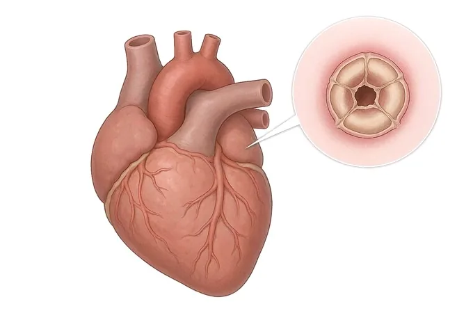
Rheumatic heart disease is not one lesion. The same immune scarring can narrow a valve, make it leak, or do both at once, and it can do this to more than one valve in the same heart. Which pattern a patient has is the most useful thing to establish, because the pattern — not the diagnosis — decides whether a valve can be repaired. The mitral valve is involved in the large majority of cases, and mitral stenosis, a valve that will not open, is the classic picture of the disease.
| Pattern | What the valve does | Why it changes the plan |
|---|---|---|
| Mitral stenosis | The leaflets thicken and their edges fuse, so the valve will not open fully; pressure builds up behind it, first in the left atrium and then in the lungs | The classic rheumatic lesion. In a suitable valve it can be opened without surgery; otherwise it is among the most repairable valves in cardiac surgery |
| Mitral regurgitation | The same scarring shortens the supporting cords and retracts the leaflets, so the valve no longer closes tightly and blood leaks backwards with every beat | Repair is usually possible and usually preferable, because a leak of this kind enlarges the heart gradually while it is tolerated |
| Aortic valve disease | The aortic leaflets thicken, fuse and retract, leaving the valve narrow, leaky, or both at once | It often means that two valves are dealt with in one operation, and it makes the choice between repair and replacement more consequential |
| Tricuspid involvement | The tricuspid valve is affected by the rheumatic process itself, or by the pressure and dilatation that a mitral lesion transmits to the right side of the heart | Adding this valve to the operation adds little to the surgery and a great deal to the years afterwards — a leak left behind here tends to return |
| Multivalve disease | Two or three valves are affected in the same heart, most often mitral together with aortic, or mitral together with tricuspid | It is the reason these operations are done in centres that do them often: the planning, the sequence and the repair techniques all change |
One framework is worth setting out here, because it organises everything on the treatment page. Where a valve can be repaired, repair is preferred to replacement. A repaired valve is the patient's own tissue, it does not carry the lifelong blood-thinning that a mechanical replacement requires, and it introduces no foreign material for infection to settle on. Replacement — mechanical or biological — becomes the right answer when the anatomy will not hold a durable repair, when several valves are badly damaged, or when a valve substitute is the safer option for that particular patient. Which of those applies is not a preference to be argued but a conclusion drawn from the echocardiogram and confirmed in the operating theatre.
How severity is read: the valve, and what it has done to the heart
Rheumatic heart disease has no stage numbers, and an echocardiogram report is not a single figure either. Severity is read on two axes together, and they are kept apart on purpose: the lesion itself — how far the valve has narrowed, or how much blood leaks backwards through it — and the consequences, meaning what that lesion has already done to the chambers behind it, to the lungs, to the rhythm and to the pump. Neither axis answers the question on its own.
| Axis | What is being read | Why it changes the timing |
|---|---|---|
| The lesion | The valve area, the pressure gradient across the valve, or the volume leaking back through it — measured on echocardiography and graded from mild to severe | It says how bad the valve is today, and repeated measurements say how fast it is changing |
| The chamber behind it | The size of the left atrium, and the size and function of the ventricle that has been carrying the extra load | A chamber that is dilating is evidence that the valve is already costing the heart something, sometimes before breathlessness is admitted to |
| The lung circulation | The pressure in the pulmonary arteries, estimated on echocardiography or measured directly at catheterisation | Pulmonary hypertension makes the operation more demanding and the recovery slower, which argues for operating before it develops rather than after |
| The rhythm | Whether atrial fibrillation has appeared, and how well the heart rate is controlled | Atrial fibrillation is common once the atrium dilates, it makes breathlessness worse, and it carries a stroke risk that needs treating in its own right |
| The pump | The ejection fraction, and whether the ventricle is coping with its load or beginning to fail | A failing ventricle changes the risk of everything, and it limits how much can safely be corrected in one operation |
Three things are worth taking from this, and none of them is about a threshold. First, the same measurement means different things in different patients: a valve area that looks unremarkable in one person may in another sit behind a hugely dilated atrium and pulmonary hypertension, and it is the combination that decides. Second, the trend replaces the photograph: a lesion that has barely moved across several yearly echocardiograms is watched, while the same lesion that has changed over two or three is discussed for surgery. Third, pregnancy changes the reading of every one of these axes, because it raises the heart's workload for months at a time — so anyone with a rheumatic valve who is planning a pregnancy is assessed before conception rather than during it, and the way that extra load brings a valve problem to light is described on the symptoms page.
What a complete assessment has to establish
The diagnosis of rheumatic heart disease is rarely a question of whether it is there. A murmur is heard, or an echocardiogram is done for another reason and the valve turns out to be abnormal — and from that point the useful questions are no longer about the presence of the disease but about what it requires.
- Which valves, and what exactly is wrong with each. Narrowed, leaking, or both — valve by valve, because a mitral stenosis and a mitral regurgitation in the same heart are two different problems needing two different repairs.
- How severe it is, measured rather than estimated. The valve area, the gradient, the volume leaking back and the pulmonary pressure, read from the images rather than inferred from how unwell the patient looks.
- What the valve has already done to the heart. Chamber sizes, pulmonary pressure, the rhythm and the pumping function — the consequences that decide the timing far more than the valve's own number does.
- Whether the rheumatic process is still active, and whether a repair is realistic. Active inflammation is treated before elective surgery; and the anatomy of the valve — the thickness and mobility of the leaflets, the state of the supporting cords, the calcium laid down — is what decides between a repair and a replacement.
Where each of those questions is answered depends on the patient: the echocardiogram answers most of them, a transoesophageal study answers the ones about a mitral valve that may be repaired, and cardiac catheterisation answers the ones on which the echo and the symptoms disagree. What matters at this level is only that the sequence, the tests and the order they come in are set out on the diagnosis page, and that the images themselves — not the written report — are what a second opinion needs.
Why patients travel to China for rheumatic valve treatment
Valve surgery travels well, and so does the decision that precedes it. Echocardiographic assessment, the classification of a valve lesion and the techniques of mitral and aortic repair are standardised internationally, and the prostheses and repair rings are the same ones used elsewhere. What separates centres is narrower and more practical: whether repair is done often enough to be offered, whether the emergency and the operating theatre sit under one roof, and how quickly a patient who is not in the country can get a written answer.
Our cardiovascular service spans two flagship hospitals — Fosun Hospital Foshan and Fosun Hospital Guangzhou — with nearly 170 specialised cardiovascular beds, modern coronary care units and advanced catheterisation laboratories. Emergency cardiac intervention runs around the clock at both. The Foshan hospital holds national certifications in five areas — chest pain, atrial fibrillation, heart failure, cardiac rehabilitation and hypertension — the first hospital in its city to do so, and the Guangzhou hospital obtained national chest pain centre certification in 2025. For valve disease that breadth matters at the margins: a patient whose irregular rhythm needs controlling, whose heart failure needs stabilising, or whose coronary arteries need assessing in the same admission is treated by teams that are on site rather than referred out.
For a visiting patient, records reviewed by a cardiology team receive a written opinion within about 48 hours of complete records arriving — and for valve disease what matters are the echocardiogram images and the measurements on them, not the summary sentence of the report. Complete treatment in China typically runs at 30–50% of comparable care in Europe or the United States, we work with more than 30 commercial insurers for direct billing, and our international patients have come from 124 countries and regions. Many patients who arrive with this diagnosis come from places where rheumatic heart disease is still the leading cause of valve disease in young and middle-aged adults, and most of them arrive already knowing that an operation is likely; what they are looking for is a team that repairs valves rather than replaces them by default. What happens to the rheumatic process after the operation is set out on the prevention page.










