


Valvular Heart Disease Overview
Valvular Heart Disease Overview
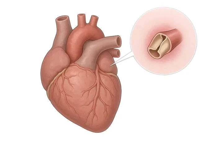
A heart valve is a one-way door, and valve disease is what happens when that door no longer opens fully, or no longer closes completely. The consequences are quiet at first: the heart compensates, the patient feels well, and the only clue may be a murmur heard through a stethoscope at an appointment that had nothing to do with the heart. This guide explains the two directions the disease can take, why a narrowed aortic valve and a leaking mitral valve behave differently, how the echocardiogram turns a murmur into a measurement, and what the timing logic of modern valve care actually is — treat when symptoms or the heart's function change, not after decompensation.
-
24/7
Emergency cover at both flagship hospitals
-
170
Specialised cardiovascular beds across the two flagship hospitals
-
5
National certifications at the Foshan hospital, the first in its city
-
124+
Countries and regions our international patients come from
What valvular heart disease is — a door that fails in one of two ways
The heart has four valves — aortic, mitral, tricuspid and pulmonary — and each is a one-way door. Blood moves through them in a single direction, and the valve's job is to open completely when the chamber behind it contracts and to close completely the moment that chamber relaxes, so that no blood travels backwards. Valve disease is the failure of one of those two halves of the job, and every symptom, every measurement and every treatment in this guide follows from which half has failed, and in which valve.
The failure has two directions. A valve that no longer opens fully is stenotic: the chamber behind it has to push blood through a smaller opening, and it does so by generating higher pressure. A valve that no longer closes completely is regurgitant, or leaking: blood that should move forward slips back, so the chamber has to pump the same volume twice. Stenosis is a pressure problem; regurgitation is a volume problem. They can feel similar to the patient, and they are measured differently.
What both have in common is the silence of the early years. The heart is an adaptable muscle: facing a narrower door, its wall thickens; facing a leaking door, its chamber enlarges. Those adaptations keep the circulation normal for a long time, which is why a person can have moderate valve disease and feel entirely well, and why the condition is often found by chance — a murmur at a routine examination, an echocardiogram done for another reason. The adaptation is also what eventually fails: a thickened wall becomes stiff and short of oxygen, an enlarged chamber loses its squeeze, and at that point the symptoms arrive together rather than gradually.
One boundary belongs here, because patients often arrive with two different diseases merged in their minds. Valve disease caused by rheumatic fever is a distinct entity with its own age pattern, its own natural history and its own guide on this site; it is not what this cluster describes, and the causes page states where the line sits. Coronary artery disease and heart failure are likewise separate conditions with separate guides. All three can coexist with valve disease — a leaking mitral valve can follow a weakened ventricle — but they are not the same problem, and their treatments are not interchangeable.
The spectrum: which valve, which direction, and what caused it
Valve disease is described by three things at once: which valve, which direction, and what caused it. Valve and direction decide the symptoms and the operative plan; the cause decides who tends to be affected and how quickly the disease moves. The table below is the practical map.
| Valve and lesion | What is happening | What it means for treatment |
|---|---|---|
| Aortic stenosis | The aortic valve, between the left ventricle and the aorta, thickens and calcifies and cannot open fully, so the ventricle pushes against a rising pressure load | The commonest valve problem in older patients. Replacement — surgical or transcatheter — is the definitive treatment, and the decision to offer it follows symptoms and the ventricle's function |
| Aortic regurgitation | The aortic valve does not seal, so blood falls back into the ventricle as it relaxes; the ventricle dilates to accommodate the extra volume | Often tolerated for years. Operation is offered when symptoms appear or when the ventricle dilates or weakens past the accepted thresholds, because after that point recovery is incomplete |
| Mitral stenosis | The mitral valve, between the left atrium and left ventricle, is narrowed — most often after rheumatic fever — and pressure builds backwards into the lungs | Breathlessness and an irregular rhythm are the usual presentation. Treatment is by catheter balloon, repair or replacement, and the rheumatic form is covered by its own guide |
| Mitral regurgitation | The mitral valve leaks as the ventricle contracts; the leak may come from the leaflets themselves, or from a ventricle that has changed shape and pulled them apart | Repair is preferred over replacement whenever the valve's anatomy allows it. Timing is the whole question: act on the valve before the ventricle is permanently affected |
| Right-sided valves | The tricuspid valve leaks as a consequence of pressure or volume problems elsewhere, most often on the left side; pulmonary valve disease is usually present from birth | A right-sided leak is usually treated by correcting what caused it; the tricuspid valve itself is repaired or replaced during the same operation when the leak is severe |
| Multiple valves | More than one valve is affected — aortic stenosis together with mitral regurgitation, or a rheumatic process that has left several valves scarred | The plan is made as one operation rather than two decisions, and the combined risk of surgery is weighed explicitly against the risk of doing nothing |
Two practical notes follow from this map. First, the same grade carries different weight in different valves: a severely narrowed aortic valve is a problem that eventually forces a decision, while a severely leaking mitral valve may reasonably be watched for years if the ventricle is coping and the patient is well. Second, cause and direction interact. A leak produced by a ventricle that has changed shape after a heart attack behaves differently from a leak produced by a torn leaflet, and the second is far more often repairable — which is why the echocardiogram is asked to describe the mechanism, not only the size.
Two axes: what the valve measures, and what it has done to the heart
Valve disease has no cancer-style stage numbers, and patients are often surprised by that. Severity is read on two axes that are kept deliberately separate. One describes the valve itself: how narrow or how leaky, measured by echocardiography and graded mild, moderate or severe. The other describes what the valve has done to the heart and to the patient — whether the ventricle is thickening, dilating or weakening, whether pressures behind the valve are rising, whether the rhythm has changed, and what the patient can still do.
| Axis | What is measured | Why it matters |
|---|---|---|
| The valve itself | How narrow or how leaky the opening is, by Doppler echocardiography and graded as mild, moderate or severe; the pressure gradient across a narrowed valve; the size and direction of a leak | The grade sets how closely the valve is watched and whether a procedure is on the table — but it does not, by itself, say when |
| The ventricle's response | Wall thickness, chamber size and pumping function, together with the size of the left atrium and the pressure in the lungs | This is the axis that decides timing. A valve that has begun to damage the ventricle is a valve to treat, even before the patient feels much |
| The rhythm | Whether an irregular rhythm has appeared and how well it is controlled; the conduction of the heart's electrical signal, which the same disease process can disturb | A new irregular rhythm in valve disease is not a side issue: it changes symptoms, stroke risk and the timing of any operation |
| The patient | Exercise capacity, breathlessness on effort, dizziness or fainting, chest discomfort, and what the patient has quietly stopped doing because of them | The patient's axis outranks the numbers. Once symptoms appear in significant valve disease, the risk of waiting begins to rise |
Two rules keep the axes from being read as one another. The first is that a grade is not a timetable: severe on a report means the valve meets a measured threshold, not that an operation is due next month, and a patient with a severe leak and a normal ventricle may sensibly be watched. The second is that the numbers must be read beside the person. The same measurements in a fit patient of eighty and in a patient with weak kidneys, lung disease or a difficult chest lead to different advice, because each option's durability has to be weighed against the years it will need to work. What is not negotiable is the direction of the logic: intervention is offered when symptoms appear or when the heart's function begins to change — not after decompensation, when the operation is riskier and the recovery less complete. The options themselves are set out on the treatment page.
How the question moves from a murmur to a plan
A valve assessment follows a fixed order, and the order exists because each step narrows the question rather than answering everything at once. The stethoscope raises a suspicion; the echocardiogram turns it into a measurement; the remaining tests answer what the echocardiogram cannot. Knowing the sequence tells a patient what each appointment is for — and why a reassuring report from last year may still need repeating.
A murmur is turbulent blood where there should be silence. It says that a valve is abnormal, roughly which one, and roughly in which direction. It does not say how severe, and it cannot see the heart's response. A murmur is the reason to look, not the answer.
Transthoracic echocardiography measures the opening, the leak, the pressures and the ventricle's function, without radiation and without pain. It is the verdict on severity, and it is repeated over time because the trend matters more than any single reading.
Depending on what the echo shows, the next tests may be a transoesophageal study for a clearer view of the valve, a stress test to see what happens under load, a CT scan for the anatomy of the aortic root, or a catheter study of the coronary arteries before an operation.
The decision is made in a valve clinic, where the measurements, the symptoms and the patient's other conditions are read together. This is the step at which most disagreements are resolved — and the step at which a second opinion is most useful.
What the assessment produces is a document, and it ought to be readable by the person it describes. A complete valve report states which valve is affected and in which direction, how severe the lesion is by the accepted measurements, what the ventricle and the atrium are doing in response, whether the rhythm has changed, and what the team proposes next — and if the answer is "watch, and repeat the echo in a year", that is a plan too, written down with its reasons. A report that says only "moderate mitral regurgitation" leaves every decision in this guide unmade.
Why patients travel to China for valve treatment
Valve disease is decided from images and measurements, and those travel well. The severity grading systems are international, the prostheses and catheter devices used here are the same ones used elsewhere, and the operations — repair, replacement and the transcatheter procedures — are standardised. What separates providers is the span of the service: whether the imaging, the operating theatre, the catheter laboratory and the rehabilitation programme sit in one place, and how quickly a visiting patient moves from records to a written plan.
Our cardiovascular service spans two flagship hospitals — Fosun Hospital Foshan and Fosun Hospital Guangzhou — with nearly 170 specialised cardiovascular beds, modern cardiac care units, echocardiography laboratories and catheterisation facilities, and cardiac surgery on site. Emergency cover runs around the clock, which matters in valve disease for the patient who has decompensated and for the infection that lands on a damaged valve. The Foshan hospital holds national certifications in five areas — chest pain, atrial fibrillation, heart failure, cardiac rehabilitation and hypertension — the first hospital in its city to do so, and the Guangzhou hospital obtained national chest pain centre certification in 2025. The teams work across the whole spectrum rather than one corner of it: valve repair rather than replacement wherever the anatomy allows, aortic and mitral surgery with on-site surgical backup, and transcatheter aortic valve implantation together with transcatheter edge-to-edge mitral repair for patients whom open surgery would treat badly.
For a visiting patient, records reviewed by a cardiology and cardiac surgery team receive a written opinion within about 48 hours of complete records arriving — the echocardiogram images themselves, not only the report, are what the review needs. Complete treatment in China typically runs at 30–50% of comparable care in Europe or the United States, we work with more than 30 commercial insurers for direct billing, and our international patients have come from 124 countries and regions. Cardiac rehabilitation, which decides how much of the result holds over the years, is delivered through a nationally certified programme rather than left to a leaflet.










