


Melanoma Overview
Melanoma Overview
Melanoma is a cancer that begins in melanocytes, the cells that make the pigment in the skin. Most melanomas start in the skin; a minority begin inside the eye or on a mucosal surface, and those behave differently because their cause is different. What makes melanoma unlike most cancers is that it usually announces itself on the outside — as a spot that changes — which is why the whole disease turns on whether a change is noticed and examined.
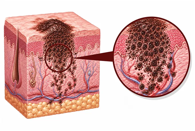
-
Depth decides most of it
Breslow thickness in millimetres, ulceration and mitotic rate — not the size you see
-
Not all melanomas are sun-related
acral, mucosal and eye melanomas arise without ultraviolet exposure
-
Check the nodes when thickness warrants
sentinel lymph node biopsy stages the first draining node
-
48 hrs
from complete records to a personalized MDT plan
What melanoma is
Melanoma is a cancer that begins in melanocytes — the cells that produce the pigment in the skin. They exist for a purpose: they sit in the deepest layer of the epidermis, and when ultraviolet light reaches them they make more pigment, which is what a tan is. It is also why a melanoma is usually a pigmented lesion. But not always: a melanoma can be pink, red or flesh-coloured, and one that has lost its pigment entirely is the kind most easily mistaken for something harmless.
Most melanomas begin on the skin, and a minority begin elsewhere. The skin is by far the commonest site, which is why the disease is described as a skin cancer and why sun protection matters. But melanocytes also exist inside the eye and in the mucosa that lines the nose, mouth and bowel, and melanomas do arise in those places. The distinction is not bookkeeping: the melanomas that arise on skin exposed to the sun are driven by ultraviolet radiation, whereas acral melanomas on the sole or under a nail, mucosal melanomas, and melanomas inside the eye arise without ultraviolet exposure and have their own biology and their own tissue markers. This is the single most useful fact for anyone of Asian or African descent, in whom those non-sun-related types are the ones that occur more often.
What makes melanoma different from most cancers is that it announces itself on the outside. A melanoma in the skin can be seen, measured and photographed, and the change that matters usually happens slowly enough to be noticed. That is the reason the disease is described in terms of a mole that changes rather than a lump that grows: the chance to treat a melanoma while it is still confined to the epidermis, or only a fraction of a millimetre deep, comes from someone looking at their own skin. The corollary is equally true — a melanoma that is dismissed as a harmless mole, a bruise on the sole or a fungal nail infection is a melanoma that has had time to thicken.
Two measurements decide what happens after the diagnosis. The first is Breslow thickness, how far the melanoma has grown down from the surface, in millimetres, measured by the pathologist. The second is whether the surface of the melanoma has ulcerated, since ulceration means the melanoma is behaving more aggressively than its thickness alone would suggest. Together with the number of dividing cells and whether the melanoma has been completely removed, these are the findings that separate a melanoma treated by complete removal alone from one that needs a check on the lymph nodes, further treatment after surgery, or a closer schedule of follow-up.
And melanoma is treatable, which is worth saying plainly. A melanoma confined to the skin is a surgical disease: the lesion and a margin of normal skin around it are removed, and that is the treatment. When a melanoma is thicker, the lymph nodes are staged and the tissue markers are read, because treatment for melanoma that has reached the nodes or spread beyond them is now built on immune checkpoint inhibitors and on targeted drugs rather than on chemotherapy — and those choices depend on tests done on tissue that already exists. The practical consequence is that the pathology report, rather than the appearance of the spot, is the document that governs the treatment.
Types of melanoma, and why the site matters
Melanomas are classified by how they behave in the skin and by where they start. The classification matters for two practical reasons: it explains why some melanomas are found early and others late, and it separates the melanomas that are related to ultraviolet exposure from those that are not.
| Type | Where it starts, and how it behaves |
|---|---|
| Superficial spreading | The commonest type on skin exposed to the sun; it grows sideways within the epidermis for a period before growing downwards, which is why it can be caught early |
| Nodular | Grows downwards from the outset, so it thickens before it is noticed; it may have little or no pigment and appears as a firm, growing lump |
| Lentigo maligna | Appears on skin that has been damaged by the sun over decades, most often on the face of older people, as a slowly enlarging flat brown patch |
| Acral lentiginous | Arises on the sole, the palm or under a nail, where ultraviolet exposure plays no part; it is the commonest type in Asian and darker-skinned populations and is often found late because of its site |
| Mucosal | Arises on the lining of the nose, mouth, anus or genital tract, again without any relation to sunlight, and tends to be found late because it is not visible |
| Uveal (eye) | Arises inside the eye, most often in the choroid, without any relation to sunlight; it may be found during a routine eye check and it has its own tissue markers |
Two points are worth carrying away from this. First, which type is commonest depends on the population: in people with fair skin, melanoma most often arises at sites of intermittent intense sun exposure — the back, the shoulders, the lower leg — whereas in people with darker skin it arises more often on the sole, under a nail or on a mucosal surface. Second, the site decides how easily it is seen: a lesion on the forearm gets noticed, one on the sole is attributed to long hours on the feet, one under a nail is treated as a fungal infection, and one inside the nose is not seen at all. That difference is why acral melanoma is so often diagnosed late.
Stage: thickness, nodes and spread
Melanoma is staged with the TNM system, and the letter that carries most weight is T — the thickness of the melanoma and whether its surface has ulcerated. What is unusual about melanoma is that the primary tumour itself is measurable to a fraction of a millimetre, which is why the pathology report is as important as the imaging.
| What is recorded | In plain terms | What it changes |
|---|---|---|
| Breslow thickness | How far the melanoma has grown down from the surface, in millimetres, measured under the microscope | Determines the margin of normal skin removed around the melanoma, and whether the lymph nodes are staged |
| Ulceration | Whether the surface of the melanoma has broken down | Raises the T category for the same thickness, and it is one of the findings that makes sentinel node biopsy worth doing |
| Mitotic rate | How many cells were seen dividing in a defined area of the melanoma | Adds to the picture of how active the melanoma is, alongside thickness and ulceration |
| Regional nodes | Whether the melanoma has reached the lymph nodes that drain the area — shown by sentinel node biopsy or by a node felt on examination | Stage III; it is the finding that makes further treatment after surgery worth discussing |
| In-transit deposits | Small deposits of melanoma in the skin or under the skin between the primary site and the regional nodes | Recorded as part of the nodal stage, and treated differently from a distant spread |
| Distant spread and LDH | Whether melanoma is present in another organ, where it is, and the level of lactate dehydrogenase in the blood | Stage IV; the site of spread and the LDH level both enter the staging of advanced disease |
Melanoma that stays in the skin and melanoma that has left it are two different diseases. Stage I and stage II are confined to the skin, and the difference within those stages is thickness and ulceration, which set how wide the margin needs to be and how close the follow-up should be. Stage III means the melanoma has reached the regional lymph nodes or is present as in-transit deposits — the setting in which treatment after surgery is discussed. Stage IV means it has spread to other organs, and it is here that tissue markers and immunotherapy come into their own.
What the tissue test adds, and what it decides
In melanoma, the tissue already in the pathology laboratory is the source of the tests that guide drug treatment. Nothing further has to be taken from the patient for most of them, which matters because it means the tests can be done while the plan is being made.
| Test | What it is | What it decides |
|---|---|---|
| BRAF | A change in a gene that drives cell division, found in a substantial proportion of melanomas arising on skin exposed to the sun | Whether treatment with a BRAF inhibitor combined with a MEK inhibitor is available, both after surgery and for advanced disease |
| KIT | A change found more often in acral and mucosal melanomas than in sun-related ones | Whether a KIT-directed targeted treatment is worth considering where the melanoma has spread |
| NRAS | Another driver change in the same signalling pathway | Explains why some melanomas do not respond to BRAF treatment, and it directs the search towards immune-based treatment |
| GNAQ and GNA11 | The changes that characterise melanoma arising inside the eye | Explains why uveal melanoma is treated differently from skin melanoma and why it is managed with its own protocols |
| PD-L1 | A protein read on the melanoma tissue that indicates whether immune treatment is likely to be of use | Contributes to the choice and sequencing of immune checkpoint inhibitors |
| LDH | A blood test rather than a tissue test, reflecting how much disease is present | Part of the staging of advanced melanoma and of the assessment of how the disease is behaving |
Two things are worth taking from this table. First, the site of origin predicts which marker is more likely to be present: BRAF is commonest in melanoma arising at sun-exposed sites, KIT alterations are more frequent in the acral and mucosal types, and uveal melanoma follows its own pathway. Testing is therefore not a fixed panel but a judgement driven by where the melanoma started. Second, the result decides how immunotherapy and targeted therapy are chosen between — and in melanoma those two classes are used far more than chemotherapy.
Why patients come to China for melanoma care
Melanoma care is decided by a set of capabilities that have to be available together — and for a disease where the treatment is chosen by a tissue marker and where the follow-up lasts years, the combination matters more than any single item:
- Pathology read with the measurements that matter — Breslow thickness, ulceration, mitotic rate and margins reported in the form that treatment decisions require, with the melanoma reviewed by a dermatopathologist.
- Dermatoscopy and skin surveillance — the examination of a lesion before it is removed, and the follow-up of a person with many moles, which is a skill rather than a photograph.
- Excision and reconstruction planned together — a margin chosen by the thickness of the melanoma and a reconstruction designed for the site, which matters most for melanomas on the face, the sole or under a nail.
- Sentinel lymph node biopsy performed as standard — with the tracer, the dye and the pathology that make the result reliable, and with nodal ultrasound surveillance as an alternative where dissection is not wanted.
- Molecular testing on the tissue that already exists — BRAF and, where relevant, KIT, NRAS and the eye melanoma markers, since these decide whether targeted treatment is available.
- Immunotherapy and targeted treatment, with their complications managed — checkpoint inhibitors and BRAF plus MEK combinations given on schedule, with the team alert to the inflammatory side effects that these drugs can cause.
- Radiotherapy for the situations where it helps — stereotactic radiosurgery for a small number of brain deposits, and palliative radiotherapy for a deposit causing pain or bleeding.
- One multidisciplinary review, and one written plan — pathology, imaging, the operation and the drug treatment reviewed at the same meeting, with the plan and the surveillance schedule set out in writing.
Because melanoma surveillance continues for years after the melanoma is removed, patients who live elsewhere commonly come for the diagnosis, the operation and the start of treatment, then return home with a written schedule that a local team can follow, coming back when a specific examination or treatment is needed. The records that make that possible are the full pathology report with the thickness, ulceration, mitotic rate and margins, and any BRAF or other marker results; the images of the lesion and of the excision site; any imaging of the lymph nodes or of other organs with its reports; and the record of treatment already given. Send those, and the written assessment is returned within 48 hours of the file being complete.










