


Congenital Heart Disease Overview
Congenital Heart Disease Overview
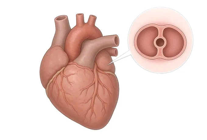
Congenital heart disease is a structural difference the heart is born with — a hole between two chambers, a valve that never opened properly, a vessel that stayed open when it should have sealed, or great arteries arriving in the wrong order. It is not one condition but a range, and the range decides almost everything: a small defect may be watched for a lifetime, while a complex one needs surgery in the first months of life and specialist care for the decades after. Most adults now living with a defect were not cured as children; they were repaired, and a repair needs watching. This guide follows one thread from end to end: what the defect is, how it announces itself, what has to be measured, and what each treatment can and cannot do.
-
24/7
Emergency cardiac intervention at both flagship hospitals
-
170
Specialised cardiovascular beds across the two flagship hospitals
-
5
National certifications at the Foshan hospital — the first in its city
-
124+
Countries and regions our international patients come from
What congenital heart disease is — a difference the heart is born with
Congenital heart disease is the name for a structural difference in the heart that is present at birth. The heart is a four-chambered pump with four valves and two great arteries, assembled in the first weeks of pregnancy from what starts as a single tube that twists and divides. Congenital heart disease means one step in that assembly did not complete, or completed in a different shape: a hole between two chambers that should have sealed, leaflets of a valve that did not separate, a duct that should have closed after birth and stayed open, or vessels that arrive in the wrong place. The defects are numerous, and they differ enormously in how much they matter — from a pinhole that never causes a problem to a defect that has to be operated on within the first days of life.
The word congenital describes timing, not severity. It says the difference was there before birth; it says nothing about the weight the patient will carry. That is decided by two other things: which structure is involved, and how much extra work the defect makes the heart and the lungs do. A defect that adds work every minute of every day for thirty years is a very different problem from one that leaves the circulation almost untouched — and the two can share the same three-letter name.
One feature of these defects explains most of their behaviour. Before birth the lungs do not do the breathing — the placenta does — so the pulmonary circulation carries little blood at low pressure, and a hole or a misplaced vessel changes very little while the baby is still inside. At birth the circulation rearranges itself within hours: the lungs open, the pressures reverse, and the defect begins to matter. From that moment the heart does what any muscle does when the workload rises — it adapts. It enlarges, thickens and keeps the circulation adequate, and it does so remarkably well for years. That adaptation is finite, and it is the reason the story of congenital heart disease so often has two chapters: a quiet childhood, and an adulthood in which the bill arrives.
Three things this guide is not about, said plainly because patients arrive with them confused. Congenital heart disease is not acquired valve disease — a valve damaged by rheumatic fever or by age is a different problem with a different repair, and the bicuspid aortic valve, the most common defect found in adults, is covered with valvular heart disease because what it eventually demands is valve surgery. It is not coronary heart disease, which concerns the arteries running on the surface of the heart rather than the structure inside it. And it is not heart failure, although a defect that has loaded the heart for decades can end there. The three can coexist in one patient; each has its own logic, and each has its own guide.
Simple and complex: the two groups that decide the pathway
Almost everything about how a defect should be managed follows from which of two groups it belongs to. Simple defects involve one structure: a hole between the upper chambers (atrial septal defect, ASD), a hole between the lower chambers (ventricular septal defect, VSD), a vessel that should have closed after birth and did not (patent ductus arteriosus, PDA), or a narrowed pulmonary valve. Complex defects involve several structures at once — tetralogy of Fallot, transposition of the great arteries, and the single-ventricle pathways — and they are named in this guide only so that a reader recognises the terms, because their care is planned by a specialist congenital team across a lifetime. The word simple describes the structure, not the stakes: a small defect can be genuinely trivial, and a simple defect first recognised in middle age can need treatment within weeks.
| Group | What is involved | What it usually means |
|---|---|---|
| Simple: a hole in the atrial septum (ASD) | An opening between the two upper chambers, letting oxygenated blood cross to the right side and back to the lungs | Often silent for decades and found in adulthood; closure is considered when the shunt is significant or the right side has enlarged |
| Simple: a hole in the ventricular septum (VSD) | An opening between the two lower chambers, the most common defect overall; small ones may close on their own in childhood | Small defects are watched; larger ones are closed to protect the lung circulation from a lifetime of extra flow |
| Simple: a persistent ductus (PDA) | The vessel connecting the aorta and the pulmonary artery, which normally seals after birth, remains open | Usually closed through a catheter without surgery, at almost any age, once the shunt is judged significant |
| Simple: a narrowed pulmonary valve | The valve between the right ventricle and the pulmonary artery does not open fully, so the ventricle works harder | Mild narrowing is watched; when the obstruction is meaningful it is relieved by balloon or surgery |
| Complex defects | Several structures are involved together — tetralogy of Fallot, transposition of the great arteries, single-ventricle pathways | Named here, not expanded: these are planned and followed by a specialist congenital team from early life onward |
The mechanism behind most of the simple defects is a shunt, and its direction gives the condition its character. In the common holes, pressure on the left side of the heart is higher, so oxygen-rich blood crosses to the right side and is pumped to the lungs a second time: the lungs receive more blood than they were built to handle. The pulmonary circulation is a low-pressure system, and it tolerates the extra volume for years — which is exactly why the defect stays quiet. But a shunt that runs for decades keeps the right side of the heart and the lung vessels under a load they were not designed to carry, and the pressure inside the pulmonary artery starts to climb. That pressure, rather than the size of the hole, is the number that decides whether closing the defect is still a good idea — and it is the reason an adult with a newly found defect is assessed so carefully before anything is done. How a shunt and lung pressure are measured is the subject of the diagnosis page.
How severity is read: the anatomy, and what the heart has already paid
Patients arrive with the same question in different words: how serious is my defect? The answer is never the name alone, and it is never the size alone. Severity is read on two axes together — what the anatomy actually is, and what the heart and the lungs have already paid for living with it — and both axes are measured rather than guessed.
| Axis | How it is measured | Why it changes the plan |
|---|---|---|
| Anatomy: which structure | Echocardiography and cardiac MRI show the defect's exact type and position, and how close it sits to valves | Position decides what can be repaired and which approach can reach it |
| Anatomy: how much blood crosses | The shunt is quantified as the ratio between the flow to the lungs and the flow to the body | A small shunt is watched; a large one is the reason to close the defect rather than continue observing |
| Cost: chamber enlargement | The size and function of the right and left chambers on imaging, and how they move | An enlarged chamber is the heart paying the bill — it is evidence the defect is no longer cost-free |
| Cost: lung artery pressure | Pulmonary artery pressure estimated on echocardiography and, when it matters, measured directly at catheterisation | Raised pressure can rule closure out altogether; it is the single measurement most likely to change the recommendation |
| Cost: rhythm and exercise capacity | Rhythm monitoring and an exercise or walking test, with oxygen saturation | Palpitations, arrhythmia and a falling exercise tolerance describe what the defect costs the patient today |
Three conclusions follow, and they are worth holding on to. First, a defect's name is not a prognosis: two patients with the same ASD may sit at opposite ends of this table. Second, lung artery pressure outranks the size of the hole — the pressure decides whether closure is still safe, so a large defect with normal pressure and a moderate defect with high pressure are managed in opposite directions. Third, the absence of symptoms is not the absence of a problem: the wear of a shunt is quiet by nature, and it is exactly the patients who feel fine whose monitoring matters most. What a defect feels like as the compensation runs out is set out on the symptoms page.
What a complete diagnosis has to answer
A diagnosis is not finished when the defect has a name. "You have an atrial septal defect" leaves every treatment decision unmade, because the question is never only which defect — it is how much blood is crossing, what the lungs have been paying, and what else the heart is doing. A complete assessment answers four questions, and all four are needed before a plan can be discussed.
| Question | The answer it needs | What depends on it |
|---|---|---|
| Which structure, and exactly where? | The type of defect, its position within the septum or the vessel, and its relationship to valves and conduction tissue | Whether a catheter device can be used at all, or whether the defect is one that surgery must reach |
| How much blood is crossing? | The shunt ratio and the measured chamber volumes, rather than a visual impression of the hole | Whether the defect is watched or closed — a decision that turns on flow, not on size |
| What have the lungs been paying? | Pulmonary artery pressure, and whether the lung vessels still respond when flow changes | Whether closure is still safe, and how urgently the assessment has to proceed |
| What else is the heart doing? | Rhythm and conduction, valve competence, pumping function and the size of each chamber | Which follow-up is needed, what can safely wait, and which complications are being watched for |
Two points of practice follow. The first is that echocardiography is the entry test in almost every case, and that the sequence after it is driven by the question the echo leaves open — MRI when the anatomy or the chamber volumes must be exact, catheterisation when the pressure must be measured rather than estimated, rhythm monitoring when the story includes palpitations or fainting. The second is that a report which names the defect and stops there is not yet a diagnosis; it is a starting point that pushes the important work onto the next visit.
What to ask for: whether the shunt is significant, what the pulmonary artery pressure is, what the chamber sizes show, and what the rhythm looks like. Those four answers are what a treatment decision is made from — and they are the four things worth having in writing before you agree to anything. What each treatment can and cannot do follows from them.
Why patients travel to China for congenital heart disease
Decisions in congenital heart disease are made from images and measurements, and both travel well. Naming, imaging standards and the principles of repair are international; what separates one centre from another is the span of care — whether the imaging, the catheter laboratory, the operating theatre and the years of follow-up sit under one roof, and how quickly a patient who arrives from abroad moves from records to a plan.
Our cardiovascular service runs across two flagship hospitals — Fosun Hospital Foshan and Fosun Hospital Guangzhou — with nearly 170 specialised cardiovascular beds, modern cardiac care units and advanced catheterisation laboratories, and emergency cardiac intervention available around the clock. The Foshan hospital holds national certifications in five areas — chest pain, atrial fibrillation, heart failure, cardiac rehabilitation and hypertension — the first hospital in its city to hold all five, and the Guangzhou hospital obtained national chest pain centre certification in 2025. Within that service, structural heart work covers device closure of atrial septal defects, ventricular septal defects and patent ductus arteriosus, balloon procedures on valves, and surgical repair with on-site cardiac surgery and intensive care backing it up. For complex defects, planning is done jointly by imaging, interventional and surgical teams rather than handed from one department to the next.
For a patient coming from abroad, records reviewed by a cardiology team receive a written opinion within about 48 hours of complete records arriving — and what the review needs is the imaging itself, not only the report. Complete treatment in China typically runs at 30–50% of comparable care in Europe or the United States; we work with more than 30 commercial insurers for direct billing, and our international patients have come from 124 countries and regions. Because a defect is a lifetime matter rather than a single episode, patients leave with a written surveillance plan — what is measured, how often, and what would change the plan. The practical questions — cost, records, travel and follow-up after you go home are answered in full on the FAQ page.










