


Carotid & Intracranial Stenosis Overview
Carotid & Intracranial Stenosis Overview
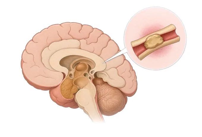
Carotid and intracranial stenosis are the same process in two places: atherosclerotic plaque building up inside an artery that supplies the brain, narrowing the channel and giving off material that travels downstream. The two places are not the same decision. At the carotid bifurcation in the neck, opening or removing the narrowing reduces the risk of a further stroke in patients who have had symptoms from it, on top of medical therapy. Inside the skull, the trials pointed the other way: intensive medical therapy alone did better than stenting, because the procedures carried a risk of stroke and death around the time they were done that outweighed the benefit they produced.
-
National
Stroke Center construction unit, and a national GCP base
-
3D DSA
Alongside 3.0T MRI, dual-source CT, PET-CT and 24-hour video EEG
-
48 hours
Written second-opinion report after complete records arrive
-
124+
Countries and regions our international patients come from
What is narrowing, and where
Atherosclerosis builds a plaque inside the wall of an artery. It thickens the wall and narrows the channel, and it does a second thing that matters just as much: the surface can become irregular and shed material downstream. Strokes from a narrowed artery happen in both ways — by flow falling below what the tissue needs at the far end, and by a fragment of plaque travelling until it blocks a smaller vessel. A narrowing therefore has two consequences that do not track each other, and a patient can have one without the other.
The carotid here means the common carotid as it divides in the neck into the internal and external branches. The plaque sits at or near that bifurcation, in the first few centimetres of the internal carotid artery. It is reachable surgically, which is part of why the evidence for treating it developed early. Symptoms from it occur on the opposite side of the body, and in the eye on the same side, because of where the artery goes.
The intracranial arteries are the ones inside the skull: the internal carotid after it enters, the middle and anterior cerebral arteries, and the vertebral and basilar arteries at the back. These vessels are smaller, they run in a confined space, and the branches coming off them supply deep structures with little alternative supply. Plaque here is harder to reach and the procedures carry more risk. It is also, in East Asian, South Asian, African and Hispanic populations, a larger share of the stroke burden than carotid disease is — the reverse of the pattern described in European populations.
What is being described on this page is atherosclerotic narrowing. Two other conditions narrow these arteries and are mentioned here only so they can be recognised and sent elsewhere: moyamoya disease, a progressive non-atherosclerotic closure of the terminal carotid with a collateral network, and arterial dissection, a tear in the wall that is a different illness with a different time course.
Carotid or intracranial, symptomatic or silent
A stenosis report is read along three axes at once, and each one changes what is offered.
Where it is. Carotid or intracranial, and within the intracranial group, anterior circulation — the intracranial carotid and the middle cerebral artery — or posterior circulation, the vertebral and basilar arteries. This axis comes first because it sets which evidence applies, and it is the one that decides whether the discussion is about opening the vessel or about medical therapy.
Has it caused a symptom. A narrowing that has produced a transient attack, a visual loss or a stroke in the territory it supplies is symptomatic. One found on a scan or heard as a bruit in a person who has never had a symptom is asymptomatic. This is the single most important distinction in the whole condition, and it is not the same thing as severity: a severe narrowing that has never caused a symptom and a moderate one that has are managed differently. The same artery can also be incidentally found in a patient whose stroke was caused by something else, in which case it is asymptomatic even though the patient has had a stroke — a distinction that is easy to lose and that changes the plan.
How much of the vessel is narrowed, and by what. Degree, measured as a percentage and meaningless without the method; and composition, whether the plaque is smooth or ulcerated, calcified or containing haemorrhage. Two narrowings of the same degree are not the same finding when the plaques differ, and this is why the report you want describes the plaque and not only the percentage.
How much, and how it was measured
Two kinds of number are being discussed, and confusing them is the most common error in a cross-border opinion. One is anatomy: how far the channel has been narrowed. The other is consequence: what the narrowing has already cost and how close to the limit the circulation is running. A patient can have a severe narrowing and never have had an event, and a patient can have a moderate narrowing that has already caused two.
| Axis | How it is described |
|---|---|
| Where the narrowing sits | Carotid bifurcation, intracranial carotid, middle cerebral artery, or the vertebral and basilar circulation. This is the axis that sets which body of evidence applies, and it comes before the percentage rather than after it. |
| The percentage, and the method used to obtain it | The two long-standing methods measure against different denominators, so the same vessel yields a higher number by one than by the other. A report that gives a percentage without saying how it was measured cannot be compared with a threshold from elsewhere. Ultrasound, CT angiography and catheter angiography each also have their own habits, which is why a change between two reports is only a change if both used the same method. |
| Whether it has caused a symptom | Symptomatic or asymptomatic, in the territory that the vessel supplies. A narrowing found after a stroke caused by something else is asymptomatic, and this is the distinction most often lost in a referral letter. |
| What the plaque is made of | Smooth or ulcerated, calcified or soft, with or without haemorrhage inside it. Ulceration and intraplaque haemorrhage are described as carrying more risk than a smooth plaque at the same degree of narrowing, which is why imaging of the plaque itself is part of a complete workup rather than an addition to it. |
| What it has already cost in flow | Whether the territory downstream is still receiving enough, whether there is a border zone pattern of injury, and whether the collateral circulation is carrying the load. Measured with perfusion imaging, and described separately from the degree — a severe narrowing with good collaterals and a moderate one with none are not equivalent. |
| How long since the event, and how complete the medical therapy is | The risk carried by a symptomatic narrowing is highest in the days and weeks after the event, which is why symptomatic carotid surgery is done early rather than scheduled. Whether the patient is already on a statin at target, an antiplatelet, and treated blood pressure is asked before anything else is offered, because intervention is added on top of that rather than instead of it. |
| What the other vessels are doing | Disease on the other side, disease in series in the same vessel, and disease in the coronary circulation. A patient with a carotid narrowing has a vascular condition and not only a neck finding, and the other territories influence both the risk of intervention and what follow-up is arranged. |
Read together, the axes do not produce a severity score. They answer which of three situations this is: a symptomatic narrowing in a territory that has declared itself, where the question is whether and when to open it; an asymptomatic narrowing found on a scan, where the question is how complete the medical therapy is and what follow-up will catch a change; or an intracranial narrowing, where the question is almost always whether the medical therapy is being delivered properly, because that is what the trials changed.
How a stenosis is demonstrated
The sequence begins with what is quick and non-invasive and proceeds to catheter angiography only when the answer would change what is done. In most patients the decisive study is not the one that measures the narrowing, but the one that shows what the narrowing has already cost.
| Investigation | What it settles |
|---|---|
| Carotid duplex ultrasound | Whether there is a narrowing, where along the vessel it sits, and how fast the blood is moving through it, from which the degree is derived. It is quick, repeatable, and the study used for follow-up precisely because it can be repeated without cost to the patient. |
| MRI of the brain with MR angiography | Whether tissue has already been lost, and in what pattern — a single territory, several, or the border-zone pattern that points to flow rather than to emboli. This is the study that tells you whether the narrowing has already cost something, and it is the one most likely to be missing from a workup done for a different reason. |
| CT angiography | The lumen, the plaque, and the arch and the vessels leading up to it, which matters if an endovascular procedure is being considered. It is fast, which is why it is used in the acute setting, and it shows calcification well. |
| Digital subtraction angiography | The reference study, which shows the vessels filling in sequence, the collateral circulation, and whether the narrowing is one lesion or several in series. It is done by the team that would perform the procedure, because the images are the plan rather than a report about one. |
| Perfusion imaging, and imaging of the plaque | Perfusion studies measure what the narrowing has cost in flow rather than how many millimetres it is. Plaque imaging — for ulceration, haemorrhage inside the plaque, and a lipid-rich core — describes the lesion rather than the channel, and is what separates two narrowings of the same percentage. |
A report worth having therefore answers six things: where the narrowing is, what percentage and by which method, whether the plaque is smooth or ulcerated, whether the patient has had a symptom from it and in which territory, whether tissue has already been lost downstream, and what the other vessels are doing. The second of those is the one most often given without its method, and the fifth is the one most often absent.
Why patients travel to China for stenosis care
A patient who is having a transient attack, or who has just had a stroke, needs care where they are, and no one should travel during an event. Travel becomes reasonable between events, and in three situations: when a narrowing has been found and the question is whether it is the one causing the symptoms and whether it should be opened; when a patient has been offered an intervention and wants a second reading of the imaging and of how complete the medical therapy is; and when the need is rehabilitation after a stroke that has already happened.
Our neurology and neurosurgery services are organised as a standardised stroke centre built to national stroke-centre standards, and are a National Stroke Center construction unit and national GCP base. Cerebrovascular cases are planned jointly by neurology, neurosurgery and rehabilitation rather than handed from one department to the next, and interventional work — carotid and intracranial stenosis, moyamoya disease, aneurysms and arteriovenous malformations — is carried out on 3D DSA, alongside 3.0T MRI, dual-source CT, PET-CT and 24-hour video EEG.
The practical questions are answered the same way as for any other condition we treat. Records reviewed by a neurovascular specialist receive a written opinion within about 48 hours of complete records arriving, and the opinion states where the narrowing is and how much, whether the imaging supports that it caused the symptoms, what the medical therapy should be and what it currently is, whether intervention is being added and by which route, and what remains uncertain. Complete treatment in China typically runs at 30–50% of comparable care in Europe or the United States. We work with more than 30 commercial insurers for direct billing and have received patients from 124 countries and regions.










