


Vestibular Schwannoma Overview
Vestibular Schwannoma Overview
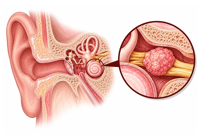
A vestibular schwannoma — still widely called an acoustic neuroma — does not arise from the brain. It arises from the Schwann cells that sheathe the vestibular half of the eighth cranial nerve, the nerve that carries hearing and balance, where that nerve runs through a narrow canal in the bone of the skull base. It is benign: it does not spread, and many of these tumours grow slowly or not at all. What makes it a distinct problem is where it lives — a bony passage with no room to spare — because a tumour there compresses the very fibres that carry sound, which is why a one-sided, gradually progressive hearing loss is usually the first thing to appear.
-
Benign
it does not spread through the body; the risks come from pressure in a narrow space
-
Hearing first
a one-sided, progressive hearing loss is the commonest first sign
-
Three findings
size, growth on successive scans, and the hearing on that side decide the plan
-
48 hrs
from complete records to a personalized MDT plan
What a vestibular schwannoma is
The nerve of hearing and balance — the eighth cranial nerve — has two halves: the cochlear half, which carries sound, and the vestibular half, which carries signals about head position and movement. Like the nerves of the body, it is insulated by cells called Schwann cells. A vestibular schwannoma is a benign overgrowth of those cells on the vestibular half. It usually begins at the inner end of the bony canal that carries the nerve from the inner ear towards the brain, and it grows from there into the space at the side of the brainstem called the cerebellopontine angle.
Three features of that location explain almost everything about the condition. The first is that the canal leaves no room to spare. A tumour growing inside it presses on the nerve fibres travelling alongside it long before it presses on the brain, and the fibres that fail first are the ones that carry sound. This is why a hearing loss in one ear, creeping up over months or years, is the usual first sign — and why the loss is often noticed on the telephone, or when one ear is blocked, rather than in a doctor's office.
The second is that it is a tumour of the nerve, not of the brain. It is benign, it does not invade the brain tissue, and it does not spread anywhere else in the body. The problems it causes are problems of compression — of the hearing nerve first, then of the facial nerve which runs through the same canal, then of the nerve of facial sensation, and only in large tumours of the brainstem itself. That is also why treatment is judged by what it protects: the facial nerve, the hearing that remains, and the structures of the brainstem.
The third is that its growth is slow and, in any individual, unpredictable. Some tumours never grow measurably; some grow a few millimetres over years; a smaller share grows faster. No test performed at diagnosis predicts with certainty which will do which — which is why observation with scheduled scans is a real treatment option here rather than a delay, and why any comparison of scans is only as good as the earlier images.
What kind of tumour it is, and where it grows
Vestibular schwannomas are described along three axes, and a patient will usually be told all three:
- By nature. It is a benign tumour of Schwann cells — in WHO terms a grade 1 schwannoma — and malignant change is exceedingly rare. The clinical question is never whether it is a cancer but what it is pressing on and whether it is growing.
- By number and origin. The great majority are single and sporadic — occurring by chance, with no inherited cause and no implication for relatives. Bilateral tumours — one on each hearing nerve — are the hallmark of the inherited condition neurofibromatosis type 2, and their discovery sets in motion a genetic assessment and a different, lifelong surveillance programme.
- By position — contained in the canal. An intracanalicular tumour lies entirely within the internal auditory canal. It is the earliest stage, often the smallest, and the one in which hearing loss or one-sided tinnitus may be the only finding.
- By position — extending into the posterior fossa. As the tumour enlarges it passes out of the canal into the cerebellopontine angle, where it may press on the brainstem, the facial nerve, the nerve of facial sensation, and eventually the pathways of the cerebellum. Position beyond the canal is what brings imbalance, facial numbness and, in the largest tumours, headache and raised pressure.
- By the tissue itself. No biopsy is needed — the diagnosis is made on MRI. Tissue is examined in the uncommon event that the tumour is removed as treatment, and it confirms the diagnosis rather than revealing a grade that changes the plan.
Size, growth and hearing classification
This tumour is not staged in the way cancers are. Three measures do the work instead — how large the tumour is, whether it is growing, and what the hearing is doing:
| Measure | In plain terms | How it is used |
|---|---|---|
| Intracanalicular | The tumour lies entirely within the canal of the hearing nerve (often reported as Koos grade I) | Symptoms are usually confined to the ear; both observation and focused radiation are common choices |
| Small | The tumour has begun to enter the space beside the brainstem but remains small (Koos grade II) | All three options — observation, radiation, surgery — are usually still on the table |
| Medium | The tumour occupies the cerebellopontine angle and may indent the brainstem (Koos grade III) | Growth and symptoms push towards treatment; hearing preservation becomes less likely to succeed |
| Large | The tumour compresses the brainstem or cerebellum (Koos grade IV) | Treatment rather than observation; the facial nerve and the brainstem dominate the planning |
| Growth | The change measured between successive scans, compared like with like | The single most influential finding for a tumour that is causing little: quiet tumours can be watched, growing tumours move towards treatment |
| Hearing class | What the affected ear can still do — how loud sounds must be, and how clearly words are understood | Shapes the choice between approaches; hearing that is still useful changes what is worth attempting |
The practical point is that size alone decides less than it appears to. A small tumour that is growing in a patient with useful hearing is treated, and a larger tumour that has been quiet for years in a patient with no useful hearing on that side may legitimately be watched. The three measures are read together, and the patient's own priorities — which hearing means what to them, and what they are willing to trade — belong in that reading.
The genetic findings, and when they matter
Molecular and genetic testing here is not about choosing a drug. It is about recognising the uncommon patient whose tumour is part of an inherited condition, because that changes the follow-up for life:
| Finding | What it is | How it changes management |
|---|---|---|
| NF2 alteration | A change in the NF2 gene, which produces a protein called merlin that regulates these cells; found in the tumour cells of schwannomas generally | When it reflects the inherited condition, it sets a lifelong surveillance programme for both sides and for the rest of the nervous system |
| Bilateral tumours | A schwannoma on each hearing nerve | The hallmark of neurofibromatosis type 2; genetic assessment is offered rather than optional |
| Young age or family history | A single tumour at an unusually young age, or schwannomas in relatives | A reasonable reason to ask for genetic assessment, since inherited forms announce themselves early |
| Other nerve schwannomas | Schwannomas elsewhere in the body, particularly multiple ones | Raises the question of the rarer schwannomatosis conditions, which have their own genetic assessment |
| Sporadic unilateral tumour | A single tumour in an adult in midlife with no family history | Genetic testing rarely changes anything; its absence from the plan is not a gap |
Two conclusions follow. First, for the great majority — a single tumour in an adult in midlife — no genetic test is needed and none changes the plan. Second, in the situations listed above, testing is worth doing properly, because a diagnosis of neurofibromatosis type 2 brings with it scheduled imaging of both sides, surveillance of the spine and other nerves, and information that matters to relatives. Asking which situation you are in is a reasonable question to put to the team.
Why patients come to China for vestibular schwannoma care
The care of a vestibular schwannoma is decided by a small number of capabilities that have to work together, and the balance between them is what the treatment consists of: a decision to treat or to watch, a choice between radiation and surgery, and the protection of the facial nerve and the hearing throughout.
- Imaging on the right protocol — thin-slice contrast MRI through the internal auditory canal, since small intracanalicular tumours are easily missed on a routine brain study, and comparison with earlier scans is what the whole decision rests on.
- Audiology that measures what matters — pure tone and speech discrimination testing on both sides, recorded before treatment, because the hearing on the affected side is one of the three findings that decides the plan.
- Precision radiation when the tumour is suitable — stereotactic radiosurgery delivered in a single session for small and medium tumours, with the head held exactly still and the dose shaped to the tumour.
- Surgery planned around the facial nerve — skull base approaches chosen by size and by hearing status, with monitoring of the facial nerve throughout the operation and the aim of removing the tumour while preserving movement.
- Hearing and balance rehabilitation — devices and strategies for one-sided hearing loss, tinnitus management and balance retraining, which are treatment in their own right rather than afterthoughts.
- One written plan, reviewed together — neurosurgery, radiation oncology and audiology reading the same scans and the same audiograms, so that the three findings are weighed once rather than argued between departments.
Because the treatment of this tumour is often a decision followed by years of scheduled scans rather than a single episode, patients who live elsewhere commonly have the diagnosis reviewed and the radiation or the operation performed in China, then return home with a written schedule for the imaging and the hearing tests that follow. The records that make this workable are the MRI images and reports with dates — every scan, not only the latest — the audiograms on both sides, the operative or radiotherapy record if treatment has been given, and the current medication. Send those, and the written assessment is returned within 48 hours of the file being complete.










