


Uterine Fibroids Overview
Uterine Fibroids Overview
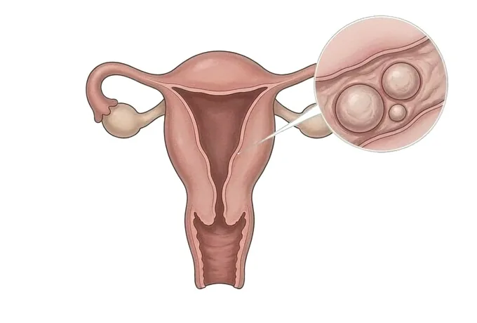
Uterine fibroids are benign tumours of the muscle of the womb. The word tumour is accurate and it is also misleading, because a fibroid is not cancer and does not turn into cancer, and most of them are found in women who have no symptoms at all. What makes a fibroid matter is where it sits. A fibroid growing into the cavity of the womb can be small and still cause bleeding that dominates a life, because it distorts the surface that bleeds. A fibroid growing outwards into the pelvis can become very large and cause no bleeding at all, only pressure. The two symptoms that follow from this are a change in the bleeding and a change in what the growing womb presses on, and they behave differently, respond to different treatments and are worth keeping separate. Two distinctions run through everything that follows. Having fibroids and needing treatment for them are two different things, because many of them never need to be touched. And the treatment that keeps the womb is not the same as the treatment that ends the problem, because fibroids can form again after they are removed.
-
Where a fibroid sits decides what it causes
a small fibroid in the cavity can cause heavy bleeding; a large one growing outwards can cause pressure and no bleeding at all
-
Fibroids are benign and do not become cancer
but a fibroid that grows after the menopause, or a new one appearing then, is the pattern that must be assessed properly
-
Having fibroids is not the same as needing treatment
where there are no symptoms and no anaemia, watching with a defined review is the correct plan rather than an omission
-
Removing the fibroids and keeping the womb is not the same as ending the problem
new fibroids can form afterwards, and recurrence is expected rather than a sign that the operation failed
What fibroids are, and what the name gets wrong
They are benign tumours of the muscle of the womb, and both halves of that description matter:
- They are tumours of muscle, not cancer. A fibroid is a collection of muscle and fibrous tissue that has grown as a lump. It is not cancer, it does not spread, and it does not turn into cancer. The word tumour is used because it is accurate, and it is worth saying clearly at the same time that in this organ it usually describes something harmless.
- Most of them cause nothing at all. A large share of women have fibroids at some point and never know it, because a fibroid that sits quietly in the wall or on the outer surface produces no bleeding and no pressure. Finding one is a finding, not a diagnosis of a problem.
- Where it sits decides what it causes. A fibroid that projects into the cavity of the womb distorts the surface that bleeds, so a small one can cause heavy periods. A fibroid that grows outwards enlarges the womb and presses on what is next to it, so a large one can cause urinary frequency and abdominal fullness with no bleeding at all. The same disease, described by its position, produces two different problems.
- They are not the same as adenomyosis. Adenomyosis is tissue like the lining of the womb growing into the muscle wall itself, spread through it rather than gathered into a lump. The two cause overlapping symptoms, they often occur together, and they answer to different treatments: a fibroid can be shelled out while the womb is kept, and adenomyosis cannot be removed at all without removing the womb.
- Having them is not the same as needing treatment. The question is not whether fibroids are present but whether they are causing bleeding, anaemia or pressure. Where they are not, the assessment ends with a plan for what to watch and when to look again.
What the name does not mean. It does not mean cancer, and it does not mean an operation is inevitable. It does not mean fertility is over, although where a fibroid sits in relation to the cavity can affect conception and carrying a pregnancy. It does not mean that a large womb must be treated or that a small fibroid must be ignored. What it means is that benign lumps of muscle have formed inside the wall of the womb, that their position is the reason for whatever symptoms are present, and that there is a sequence of options to work through rather than a single answer.
Where they sit, and what that decides
The position of a fibroid, and how far it has grown, decides what it causes and which treatments are even possible:
| Where it sits | What follows from it |
|---|---|
| Submucous, projecting into the cavity | The type most likely to cause heavy bleeding and to affect conception, and the one where size predicts the least. A small one can dominate a life, and it is also the type that can be removed through the cervix without an abdominal incision |
| Intramural, within the wall of the womb | The commonest type. It enlarges the womb, and whether it causes bleeding depends on how close it lies to the cavity and how much of it pushes inwards. It is the type where the decision to operate is least straightforward |
| Subserosal, growing outwards into the pelvis | The type most likely to cause pressure rather than bleeding: urinary frequency, constipation, abdominal girth and backache. It can become very large before anything is noticed, and it does not usually disturb the periods |
| Pedunculated, on a stalk | A fibroid on a stalk, either outside the womb or hanging into the cavity. The stalk is what matters: a fibroid on a twisted stalk causes sudden severe pain, and one hanging into the cavity behaves like a submucous fibroid |
| Several at once, or with adenomyosis | Common, and it changes the plan because the womb has to be assessed as a whole rather than one lump at a time. Where adenomyosis is present as well, removing the fibroids will not remove the disease in the wall, and the plan has to say which of the two is being treated |
Why position is asked about first. A report that names the size of the largest fibroid without saying where it sits does not answer the question that decides treatment. A two-centimetre fibroid in the cavity and a ten-centimetre fibroid on the outer surface are three different problems, with different symptoms, different risks and different operations. Establishing the position of each one is therefore the first thing the assessment does.
What changes the plan, and what does not
What changes the plan is what the fibroids are doing, and what is wanted next — not how large the womb measures:
| What changes the plan | What it changes |
|---|---|
| Whether there are symptoms at all | Where there are none and the blood count is normal, the plan is to watch with a defined review rather than to intervene. This is a decision, not a delay |
| How much the bleeding costs in daily life | It decides whether the first step is a measure that reduces bleeding, and it is the measure the plan is reviewed against. The blood count is checked rather than assumed, because bleeding that has been tolerated for years can still have produced anaemia |
| Whether pressure rather than bleeding dominates | Urinary frequency, constipation and abdominal girth come from the size and position of the womb rather than from the bleeding, so they point at different fibroids and are treated by different means |
| Whether a pregnancy is wanted now, later, or not at all | It decides which options are on the table. Methods that suppress the cycle control the symptoms and prevent pregnancy; a myomectomy is planned around preserving fertility and leaves a scar on the womb; removal of the womb ends the question |
| Where each fibroid sits, and how many there are | It decides which operations are technically possible through the cervix and which need an abdominal approach, whether the cavity can be preserved, and how much of the womb would remain afterwards |
Why size is the wrong first question. Patients usually arrive knowing how large the largest fibroid is, and it is the number least able to answer what to do. A womb can be markedly enlarged with nothing that needs treating, and a cavity can be distorted by something small enough to be missed on a report. The useful questions are whether anything is being lost through the bleeding, what is being pressed on, and whether a pregnancy is wanted.
What the assessment is looking for
The assessment measures the bleeding, characterises the pressure, examines the womb and uses imaging for what it can show:
| What is assessed | What it establishes |
|---|---|
| The bleeding history, taken as a change over time | How the periods differ from a few years ago, how many days of heavy flow there are, whether clots are passed and whether bleeding occurs between periods, which together describe how much is being lost |
| The pressure symptoms | Urinary frequency, constipation, abdominal girth and backache, which come from the size and position of the womb and point at fibroids the bleeding history would not identify |
| A pelvic examination | The size, shape, mobility and tenderness of the womb, felt directly, which changes both what is suspected and what can be planned |
| A transvaginal ultrasound | The number, size and position of the fibroids, and at the same time the lining of the womb, the ovaries and whether the muscle itself shows the changes of adenomyosis |
| A look at the cavity where a fibroid lies near it | Saline infusion sonography or hysteroscopy, because how far a fibroid projects into the cavity decides whether heavy bleeding can be treated through the cervix rather than by an abdominal operation |
| An MRI where it will change something | The position and blood supply of each fibroid in a womb that is very large or contains several, and whether adenomyosis is present as well, used to plan an operation rather than to make the diagnosis |
Why the assessment stops when it has answered the question. Not every woman needs an MRI, and nobody needs repeated imaging to watch fibroids that imaging does not guide the treatment of. The assessment is finished when the bleeding has been measured, the pressure has been characterised, the position of each fibroid has been established, the blood count is known, and the other causes that would change the treatment have been excluded.
Why patients come to China for this assessment
This is a condition whose treatments differ in what they cost a woman, and travelling for it is usually about having the position and the options set out together:
- The position of each fibroid is established rather than the size of the largest one. Ultrasound is read for where each fibroid sits in relation to the cavity and the outer surface, and the cavity is looked at directly where that finding would change what is offered.
- The bleeding and the pressure are treated as two lines. How much is being lost, whether it has produced anaemia, and what is being pressed on are recorded separately, because the two lines answer to different treatments and one can be improving while the other is not.
- The options that keep the womb are set out alongside the one that does not. Watching with a review date, medical control of the bleeding, myomectomy, hysteroscopic resection, embolisation and removal of the womb are all discussed before a plan is written, rather than one of them being presented as the only answer.
- Fertility is treated as a factor rather than an afterthought. Whether a pregnancy is wanted is established before the plan is made, because it decides whether an operation that leaves a scar on the womb is worth undertaking at that moment.
- The plan and the records are prepared to travel. Results are issued with their dates and the technique used, and the written plan names what is being treated, what is being watched, when the next review is, and what would change the approach.
What this does not promise. A completed assessment does not guarantee that the bleeding will stop or that the pressure will resolve, and it does not promise that a pregnancy will follow. What it does is establish how much is being lost, where each fibroid sits, which options preserve the womb and which do not, and what the order of trying them should be. Saying which parts can be acted on and which cannot is part of the assessment rather than a caveat added afterwards.










