


Ureteral Stricture Overview
Ureteral Stricture Overview
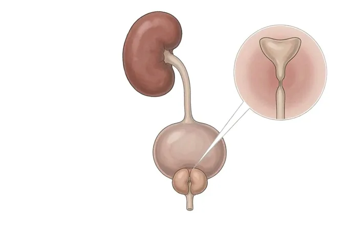
Each kidney drains through a tube about the length of a forearm. The ureter is not a passive drainpipe: its muscular wall pushes urine along in small spurts, from the kidney to the bladder, many times an hour. A ureteral stricture is a segment of that tube where the channel has narrowed — scar tissue, a congenital segment, or a piece of the tube pinched from outside — so that urine meets resistance on the way out. Everything that matters about this condition follows from the fact that a kidney does not stop working when its outlet narrows. It goes on producing urine against the blockage, pressure builds inside the collecting system, the kidney dilates, and the working tissue of the kidney is gradually squeezed and lost. That damage is usually painless. A kidney can give up much of its function without producing a symptom that would make anyone seek help, which is why a stricture is found on a scan as often as it is found by a patient. Two questions decide what happens next, and both are measured rather than assumed. Is this narrowing actually obstructing, or is it a narrow segment that the kidney empties through perfectly well? And how much function is left on that side — enough to justify rebuilding the tube, or so little that the honest answer is to leave the kidney alone? Answer those two and the choice becomes clear: a short stricture can often be relieved from inside, through the natural route, without an incision; a long, ischaemic or recurrent one is repaired with an operation that re-joins the ureter to itself or re-implants it into the bladder; and a kidney already beyond saving may legitimately be left, or removed, with the other kidney carrying the load.
-
A narrowing is not the same as an obstruction
only the functional test tells you whether the kidney is actually under pressure
-
The kidney is the casualty, not the tube
silent, cumulative loss of function is why this condition is worth treating
-
Cause changes everything
a benign scar and a tumour of the ureter look alike on a scan and are managed differently
-
48 hrs
from complete records to a personalised MDT plan
The ureter: the tube urine has to travel through
The ureter runs from the pelvis of each kidney down to the bladder — a muscular tube roughly 25 to 30 centimetres long, whose wall contracts in waves to push urine along rather than letting it fall. A stricture is a segment of that tube that has narrowed, so urine meets resistance on the way out.
It is worth being precise about what a stricture is and is not. It is not a stone, although a stone that stayed in place long enough to inflame the wall is one of its commonest causes. It is not a growth, although a tumour of the ureter itself, or a mass pressing on it from outside, produces exactly the same picture and must be excluded before anything is repaired. Mostly it is scar tissue — a segment that healed by contracting, or lost its blood supply, or is still kinked by the structure around it. None of that is a patient’s doing: the commonest cause in adult practice is a previous procedure on the ureter or in the pelvis.
The kidney is what actually suffers. A blocked drain does not stop the tap. The kidney goes on filtering, and because that urine cannot leave freely, pressure rises backwards through the renal pelvis and calyces. The collecting system dilates — this is hydronephrosis — and the same pressure is transmitted into the substance of the kidney, where it gradually thins the tissue that does the work. That is the whole argument for taking a painless narrowing seriously: the loss is silent and cumulative, and a kidney can be substantially devalued before anyone notices.
One distinction carries more weight than any other here. A narrow ureter and an obstructed ureter are not the same thing. Stricture describes the anatomy — a segment that has narrowed. Obstruction describes the function — urine failing to drain because of it. A dilated system is not automatically obstructed, and not automatically a stricture: kidneys are found dilated after a stone has passed, during pregnancy, and in wide but perfectly draining congenital systems. Only a functional test separates them, and the difference decides everything, because a narrow ureter that drains needs watching, while an obstructed one needs relieving.
Describing a stricture: cause, level, length and completeness
Urologists describe a stricture along four axes at once, and each of them constrains the options.
| Axis | What it means | Why it matters for the plan |
|---|---|---|
| Benign or malignant | Scar, ischaemia or congenital narrowing, as opposed to a tumour of the ureter wall or a tumour compressing it | The first fork in the road, taken before any repair: malignant narrowing is a different disease, and tissue diagnosis comes first |
| Intrinsic or extrinsic | Inside the wall — scar, congenital narrowing, a stone-related stricture — as opposed to a segment pinched from outside | Decides whether the tube itself must be rebuilt or simply freed from what compresses it; the two operations are not interchangeable |
| Level | Upper, at the pelviureteric junction; middle, over the pelvic brim; or lower, from the iliac vessels to the bladder | Determines which operation is anatomically possible, and how much of the native ureter can be preserved |
| Length | A short focal narrowing, typically under two centimetres, versus a long segment or one that has already recurred | Short benign strictures can often be treated from inside; long ones cannot, because the repair must bridge a gap no endoscope can close |
| Complete or partial | Whether any urine passes at all | Completeness sets the clock: complete obstruction with infection, or in a single kidney, is a same-day emergency |
A related category is obstruction at the pelviureteric junction, where the kidney meets the ureter. It may be a congenital narrowing, a segment that fails to relax, or a blood vessel crossing the junction and kinking it. It behaves like other strictures, with the same silent loss of function, but is often detected earlier because the pain is characteristically provoked by a large fluid load.
Two axes: how much the kidney has lost, and how fast
Severity here is not a grade. It is a pair of readings, and it is the pair together that tells you what to do.
Axis one is the narrowing and its effect: what level it sits at, how long the segment is, whether any urine gets past, and whether the kidney looks under pressure. Axis two is function and time: what proportion of the total kidney work that side is doing, how the system drains under a fluid load, and how long the obstruction has been present. A short narrowing with preserved function is a different problem from a long narrowing on a kidney that has already lost most of its working tissue, even when the scan appearances are equally unimpressive.
| What is measured | What it tells you | Why it matters for the plan |
|---|---|---|
| Split renal function | What proportion of the total kidney work is being done by the affected side | The reading that most changes the plan: useful function justifies reconstruction; almost none may fairly be left alone |
| Drainage on diuretic renography | Whether the dilated system empties under a fluid load, and how slowly | Separates a narrow but harmless ureter from an obstructed one — the distinction that decides whether an operation is indicated at all |
| Partial or complete obstruction | Whether any urine is getting past the narrowing | Complete obstruction with infection, or in a solitary kidney, is an emergency; partial obstruction allows a calm assessment |
| Duration and trajectory | How long the obstruction has existed, and whether function is falling on repeat testing | Recent loss can often be recovered after relief; long-standing loss frequently cannot, so a falling trend is itself a reason to act |
One caution about the numbers. Split function and drainage are reported per kidney, as a percentage and as a time, and they are read together with creatinine, which describes the two kidneys in combination. A patient can therefore have one badly affected kidney and a completely normal creatinine, because the healthy side compensates — reassuring about the body, and misleading about the disease. The affected kidney has no such spare capacity.
How a ureteral stricture is assessed
The assessment runs in a fixed order, because each step exists to answer a question the one before it cannot.
| Step | What it answers |
|---|---|
| Ultrasound of the kidneys and bladder | Is the collecting system dilated, and does the bladder empty? It raises the question, and cannot by itself separate obstruction from harmless dilatation |
| CT urography | Where the narrowing is, how long it runs, and what is causing it — a stone, a tumour of the ureter, or a mass pressing from outside |
| Diuretic renography (MAG-3) | Whether the narrowing actually obstructs, and what each kidney contributes — the functional question imaging cannot settle |
| Retrograde or antegrade pyelography | The exact calibre and length of the stricture, measured along the tube rather than estimated; drainage or a tissue sample can be taken at the same time |
| Blood and urine tests | Creatinine and eGFR for overall function; urinalysis and culture for infection and blood; cytology when a tumour is possible |
| Tissue diagnosis, when the cause is uncertain | Whether the narrowed segment is scar or tumour — answered before reconstruction is planned rather than after |
Two practical notes. Magnetic resonance urography replaces CT urography when ionising radiation is best avoided, as in pregnancy or where contrast is a problem. And a stent placed as an emergency is not part of the diagnosis: it buys time and protects the kidney while the assessment is completed, and the decision about definitive repair is taken afterwards, with the measurements in hand.
Why patients travel to China for ureteric reconstruction
Ureteric repair is a specialty of its own, and it rewards a centre that sees a steady volume of these problems. The difficult cases — strictures that have recurred after endoscopic treatment, strictures after radiotherapy, long segments where the ureter cannot simply be re-joined — are reconstructive operations, and the difference between a straightforward repair and a difficult one is made by how carefully the length, the level and the blood supply are planned for. The urology departments in our network work with the full reconstructive repertoire: ureteroureterostomy, re-implantation with a psoas hitch or a Boari flap, pyeloplasty for pelviureteric junction obstruction, and ureteric substitution using bowel or buccal mucosa for long defects, alongside the endoscopic options for short ones.
Around that sits a system built for patients who travel: imaging and renography completed quickly and read together, a report review before departure, and a written multidisciplinary plan within about 48 hours of complete records arriving. Cost belongs in the honest part of the offer — complete treatment here is typically 30%–50% of comparable care in Europe or the US, with more than 30 international insurers working with us on direct billing, and patients from 124 countries and regions. What patients tend to mention afterwards is not the operation. It is that the two questions that decide everything were answered with measurements rather than reassurance, and that when a kidney could not be saved they were told so plainly and given the reasoning.










