


Thyroid Eye Disease Overview
Thyroid Eye Disease Overview
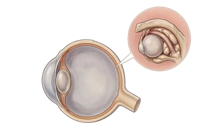
Thyroid eye disease is an autoimmune condition in which the immune system attacks the tissues behind the eye — the fat, the connective tissue and the muscles — rather than the eye itself. It is most often associated with Graves’ disease, but it keeps its own timetable: the eyes can be affected when the thyroid gland is overactive, when it is normal and when it is underactive, so ordinary thyroid blood tests never rule it out. The disease inflames and then enlarges the orbital contents. The tissues swell and the eye is pushed forward; the upper eyelid is pulled back, so the face takes on a wide-eyed look; the muscles that move the eye thicken and stiffen, which is what produces double vision; and a surface no longer properly covered becomes dry, gritty and sensitive to light. The condition moves through two phases. An active, inflammatory phase — the phase in which medication can still change the course — is followed by a quiet phase in which the inflammation settles but the changes it left do not: the bulge, the retracted lids and the double vision stay, and are corrected only by surgery. Most patients never reach the stages that threaten sight, but a small proportion develop compression of the optic nerve, which is an emergency rather than a variation. Smoking is the strongest avoidable factor of all: it makes the disease more likely, more severe and more resistant to treatment. What works is a settled thyroid, inflammation treated while it is active, a protected eye surface, no cigarettes, and any surgery planned for the quiet phase, when it will hold.
-
It is neither the thyroid gland nor the eye
the immune system attacks the fat, tissue and muscles behind the eye, and thyroid blood tests can be entirely normal
-
Two phases, two kinds of treatment
the active phase is treated with medication; the changes left behind are corrected surgically once the disease is quiet
-
Sight is threatened rarely, and urgently
most patients never lose vision, but optic nerve compression needs assessment within days
-
48 hrs
from complete records to a written assessment plan
What thyroid eye disease is, and why the thyroid is not the target
Thyroid eye disease is an autoimmune condition in which the immune system attacks the tissues of the orbit — the fat, the connective tissue and the muscles that sit behind the eye — rather than the eye itself. It is most often found alongside Graves’ disease, which is why the two names travel together, but the eye disease has an independent course: it can appear before the thyroid problem is diagnosed, at the same time, or years afterwards. Nothing about the name should suggest that the eye is simply reporting the thyroid’s state.
The mechanism explains the whole picture. Immune cells recognise something in the orbital tissues as a target and move in; the tissues swell with fluid and with long-chain sugars that hold water; the fat behind the eye increases in volume, and the muscles that move the eye thicken, first at their bellies and later with fibrous tissue. The orbit is a closed cone of bone, so enlargement has nowhere to go except forward and inward: the eye is pushed out, the muscles lose their slack and stop working together, and the eyelid is pulled back by the very tissue that has changed.
- The thyroid and the eye keep separate calendars. The eye disease can be present when thyroid function is overactive, when it is normal and when it is underactive. Normal thyroid blood tests do not exclude it, and treating the thyroid does not by itself treat the eye.
- There are two phases, and only one of them responds to medication. In the active phase the tissues are inflamed and treatment can change what happens. In the quiet phase the inflammation has settled, but the enlargement and the scar it left remain — and those are corrected with surgery, not with drugs.
- Sight is threatened in a minority, and it is an emergency when it happens. Enlarged muscles can compress the optic nerve, and an eye that cannot close can damage its own surface. Neither is common; both need to be recognised within days rather than months.
What treatment can and cannot do. Treatment can quiet the inflammation, protect the surface of the eye, relieve double vision while the disease is active and, in a minority, rescue a compressed optic nerve. What it cannot do is reverse the structural change the disease has already made. The bulge, the retracted lid and a squint that has become fixed are the province of surgery, and surgery is deliberately postponed until the disease is quiet — because operating on inflamed tissue produces results that do not hold.
The phases thyroid eye disease passes through
Thyroid eye disease is described in two dimensions at once: how active it is and how severe it has become. Severity is what the patient sees in the mirror; activity decides whether medication is offered, because immunosuppressive treatment works on inflammation and not on structure.
| What it is called | What is happening | What it means for treatment |
|---|---|---|
| Active phase | The orbital tissues are inflamed and swollen; the eye aches, the lids are puffy and the eye is being pushed forward | This is the phase medication can change. The window is measured in months, and it closes — which is why the timing of treatment matters more here than in most conditions |
| Quiet phase | The inflammation settles, but the enlarged fat, the thickened muscles and the fibrous tissue stay | Medication no longer helps. What remains — the bulge, the retracted lids, a fixed squint — is corrected with surgery, once the disease has been stable long enough for the result to hold |
| Mild disease | Lid retraction, some puffiness, dryness and grittiness, but no loss of function | Treated supportively: lubrication, protection of the surface and monitoring. Nothing more aggressive is justified by the severity alone |
| Moderate to severe disease | Marked protrusion, double vision, pain and surface damage — the changes that interfere with daily life | Immunosuppressive treatment is offered while the disease is active; radiotherapy is used in selected eyes; surgery is planned for later, when the disease has gone quiet |
| Sight-threatening disease | Compression of the optic nerve, or an eye surface that has broken down because the eye cannot close | An emergency. Treated urgently whatever the phase, because the tissue at risk does not wait for the disease to become quiet |
Why activity and severity are not the same question. An eye can be markedly protruding and completely quiet — stable for years, with nothing left to calm. The same eye can be only slightly protruding and actively inflamed, which is the situation in which medication changes the outcome. Reading only the mirror would miss the second; reading only the activity score would underestimate what surgery will eventually be asked to do.
Where the two eyes differ. The disease is usually bilateral but rarely symmetrical, and it can move at different speeds on each side. One eye may be in the active phase while the other has already settled, which is one reason the assessment is always performed and recorded for each eye separately.
What makes thyroid eye disease more or less urgent
Within any phase, a short list of findings decides how quickly things must happen. Almost all of them are established in the same assessment, and two of them are the reason a patient may be asked to return within days.
| Feature | Why it changes the plan |
|---|---|
| Whether the optic nerve is compressed | The single most serious finding in this disease. It can develop without the eye looking any different, and it is treated urgently — with high-dose treatment or decompression — because the loss it causes can become permanent |
| Whether vision or colour vision has changed | A fall in acuity, or colours looking washed out in one eye, is the earliest warning of nerve compression and is acted on straight away rather than at the next visit |
| Whether the disease is active now | Activity decides whether immunosuppressive treatment is appropriate at all. Offering it in a quiet eye treats nothing and exposes the patient to side effects for no gain |
| Whether the eye closes at night | An eye that stays open exposes its own surface to drying and ulceration. Protective measures are simple, and neglected surface damage is not |
| Whether double vision affects straight-ahead gaze | Double vision only at the extremes is tolerated while the disease runs its course; double vision in the primary position affects walking, reading and driving, and changes how quickly something must be done |
What urgent means in this disease. For most patients the timescale is months: the disease has a natural course, and treatment is planned around it. The exception is optic nerve compression, where the timescale collapses to days and sometimes hours. That asymmetry is worth carrying away from this page, because it is the one thing in thyroid eye disease that must never be watched.
How the eye and the orbit are assessed
The assessment answers four questions in order — is this thyroid eye disease, is it active, what has it done to the structure, and is sight at risk — and each step exists to make the next decision precise.
| Step | The question it answers |
|---|---|
| History and clinical pattern | Is this thyroid eye disease at all? Lid retraction, both eyes involved unevenly, and a course that has changed over months are what identify it, and the diagnosis is made from the pattern rather than from a single test |
| Measurement of protrusion and lid position | How far has the eye moved forward, and how much of the surface is uncovered? Recorded with the same instrument each time, so that the numbers can be compared rather than remembered |
| Structured activity scoring | Is the disease inflamed now? The score records the clinical signs of active inflammation and is the number that decides whether immunosuppressive treatment is offered |
| Thyroid function and antibody tests | Is there a thyroid problem to find and treat? These tests establish the association and guide thyroid management; they neither confirm nor exclude the eye disease |
| Vision, colour vision, fields and the optic nerve | Is sight at risk? Acuity, colour, pupil reactions, visual fields and examination of the nerve are recorded at every visit, with imaging added where compression is suspected or surgery is being planned |
Why the order matters. The pattern establishes the diagnosis; the measurements make the structure a number; the activity score decides whether medication has a role; and the vision assessment is the alarm that overrides everything else. An assessment that produced a bulge measurement without an activity score, or a plan without a documented visual field, would be missing precisely the information that changes what happens.
Why patients travel to China for thyroid eye disease care
Thyroid eye disease is managed over months and years, and it needs several specialties to agree with each other: ophthalmology, endocrinology, and the surgeon who will eventually operate. What an international pathway offers is that the disagreements about timing — which are the difficult part of this disease — are resolved in one place rather than across separate clinics.
The eye assessment and the thyroid picture are handled in one visit. Orbital examination, protrusion measurement, activity scoring, visual field testing and imaging are available in one facility, alongside the thyroid blood tests and the endocrine review that the eye findings must be read against. A patient leaves with one plan instead of two opinions and a question.
The decision that matters most is made with the evidence in front of it. Whether to treat with immunosuppression depends on activity; whether to operate depends on stability and on structure. Both are answered here with the measurements, the imaging and the visual fields taken during the same assessment, which is what the timing of this disease demands.
Urgent problems are treated urgently. Where the optic nerve is compressed, the pathway does not run at outpatient speed: decompression, and the intensive treatment that precedes it, are arranged without waiting for a routine slot. This is the one part of thyroid eye disease in which a day matters.
The plan is written for the patient going home. How active the disease is now, what that means for treatment and for monitoring, what the eyelids and the muscles are likely to do next, which symptoms require immediate attention, and which measurements the next treating team should inherit are all provided in English; the international desk coordinates with the patient, with any insurer or assistance company, and with the treating team at home.










