


Skin Lesions and Nevi Overview
Skin Lesions and Nevi Overview
This page covers a group of conditions that share a location rather than a cause: the marks and lumps that appear on the surface of the skin. Pigmented naevi — moles — are the most familiar, and the same group takes in seborrhoeic keratoses, skin tags and fibromas, cysts, and vascular lesions. What they have in common is that they are visible, that almost all of them are benign, and that the decision a patient is trying to make is not whether this is cancer but two separate questions, in a fixed order. The first is what the lesion is, which is settled by examining it and, where it is atypical, by taking it off whole for a pathologist to look at. The second is whether it should be touched at all — and for many lesions that are correctly identified as benign, the answer is that it should be watched rather than removed, because removal carries a scar and a recorded baseline is the safer course. The order of those two questions is what decides the outcome, and this page explains why.
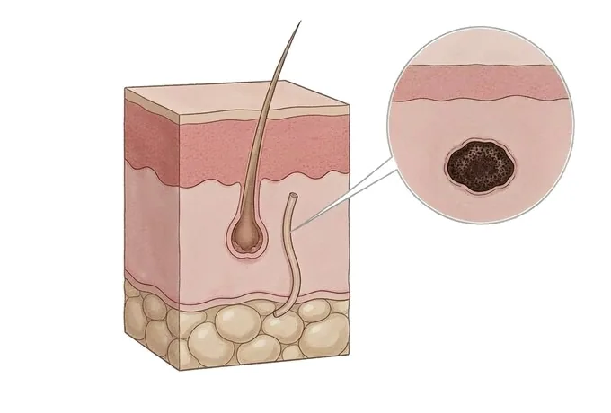
-
Moles and skin lumps are not one condition
they are a group of different benign conditions that share a location, and they are not managed in the same way
-
Almost all of them are benign
which is why the assessment is a matter of judgement rather than alarm, and why watching is often the correct answer
-
Change is the most useful observation
a lesion that has altered in shape, colour or size over months is considered differently from one that has looked the same for years
-
The order of the steps cannot be reversed
for anything atypical, the whole lesion is removed and examined rather than removed by a method that leaves nothing to examine
Why a mole and a lump are two questions, not one
This page covers a group of conditions that share a location rather than a cause: the marks and lumps that appear on the surface of the skin. Pigmented naevi — moles — are the most familiar, and the same group includes seborrhoeic keratoses, skin tags and fibromas, cysts, and vascular lesions. What they have in common is that they are visible, that almost all of them are benign, and that the decision a patient is trying to make is not whether this is cancer but two separate questions, taken in a fixed order.
- The first question is what it is. Answering it means examining the lesion rather than guessing from its appearance, and where it is atypical it means taking it off in one piece so that a pathologist can look at the tissue. Until that question is answered, no treatment is chosen.
- The second question is whether it should be touched at all. Many lesions that are correctly identified as benign are best left alone: they are not dangerous, removal carries a scar, and observation with a recorded baseline is the safer course. Being able to say that a lesion can be watched is a result, not an omission.
- The order of those two questions is what makes the difference. A lesion that is destroyed before it is identified cannot be identified afterwards, because the tissue that would have answered the question no longer exists.
Why this page states the order so plainly. A patient who has had a lesion removed with a technique that destroys it, and who then wants to know what it was, has lost the only specimen that could have said. The rule that follows is short: for anything that looks atypical or has changed, the first step is a diagnosis, and the way to obtain one is to take the lesion off whole.
The kinds of lesion this term covers
These are different conditions that behave differently, and knowing which one is being described decides whether the answer is to watch it, to treat it, or to remove it and send it for pathology.
| Kind of lesion | What it is | What it means for treatment |
|---|---|---|
| Melanocytic naevus (mole) | A cluster of pigment-producing cells, present from birth or appearing later, usually brown and uniform in colour and remaining unchanged for life | Usually observed. Removal is considered when it changes, when its appearance is atypical, or when the patient wants it gone and accepts the scar |
| Seborrhoeic keratosis | A benign overgrowth on the surface that often looks as though it is stuck onto the skin, with a waxy, scaly or crusted top | Benign, and no medical treatment is needed. It may be removed if it is repeatedly irritated, catches on clothing, or is a cosmetic concern |
| Fibroma, skin tag or cyst | Firm or soft benign lumps, including dermatofibromas, soft fibromas and epidermoid cysts, which are usually painless | Benign. Removal is offered where the lump is symptomatic, becomes inflamed or infected, or bothers the patient |
| Vascular lesion | Red or purple lesions formed of blood vessels, such as haemangiomas and telangiectasia | Managed according to the specific type. Many are benign and are simply observed; some require assessment before a decision |
| Atypical or changing lesion | A lesion that does not fit a benign pattern, or that is changing in shape, colour or size, or that bleeds, itches or crusts without a cause | The pathway is different: it is removed whole and sent for pathology, and destructive methods such as laser or freezing are not used |
Why they are listed together before anything is said about treatment. Read as one list, they suggest that a single method could serve for all of them. Read carefully, the first four are conditions that can be watched or treated at leisure, while the fifth is the one for which the order of the steps cannot be relaxed.
What makes a lesion more or less of a concern
The instinct is to judge a lesion by how alarming it looks, and that instinct is unreliable. What changes the decision is a short list of things that can be observed and recorded.
| Feature | Why it changes the plan |
|---|---|
| Change over time | A mole that has looked the same for twenty years and one that has altered in shape, colour or size over a few months are not managed in the same way. Change is the single most useful observation a patient can bring |
| Form: symmetry, border, colour, size | A lesion that is asymmetric, has an irregular or blurred border, holds more than one colour, or is larger than the patient’s other moles departs from the benign pattern and warrants examination rather than observation |
| Bleeding, ulceration or itching without a cause | A benign mole does not bleed or ulcerate by itself. These features move a lesion out of the group that can be watched and into the group that should be assessed |
| Site and repeated irritation | Lesions on the palms, the soles, the waistline or anywhere that is repeatedly rubbed or caught are considered differently, because irritation both produces symptoms and complicates follow-up |
| Number of lesions and family history | A person with many moles, or with atypical moles or a family history of skin cancer, is followed on a schedule rather than seen once |
Why "it looks bad" is not the criterion and "it has changed" is. A large, very dark mole that has not altered in two decades is usually not the one that needs attention; a small one that has changed over a few months is. This is also the observation a patient is best placed to make, which is why the history begins with it.
How a skin lesion is assessed
The assessment is built to answer the first question before any decision is taken about the second, and most of it is non-invasive.
| Step | What it establishes |
|---|---|
| History | How long the lesion has been present, whether it has changed, whether it bleeds or itches, whether it has been injured or treated before, and whether there is a personal or family history of skin cancer or of many atypical moles |
| Examination of the whole skin surface | The whole surface is examined rather than only the lesion that prompted the visit, so that other lesions are not missed and each one can be compared with the patient’s own pattern |
| Dermoscopy | A magnified view of the pattern and the distribution of pigment, which is the main non-invasive way of separating a benign lesion from one that needs to be removed |
| Photography and measurement | A photographic baseline with recorded dimensions, so that follow-up compares the lesion with its earlier state rather than with memory |
| Excision for pathology | Used where a lesion is atypical or there is reason to suspect something else. Its purpose is to provide a whole specimen to the pathologist, not simply to take the lesion off |
Why pathology is described as a diagnostic step. It answers the first question, and the answer determines what happens next. A lesion that is removed without being examined, by a method that leaves nothing to examine, has had its treatment chosen before its diagnosis was known.
Why patients travel to China for the assessment of a skin lesion
A skin lesion is a problem of judgement rather than of equipment. The difficulty is not removing a lesion but deciding which ones should be removed, which can be watched, and which need to be examined under a microscope before anything else happens. A pathway that brings assessment, dermoscopy, excision and pathology into one place is what makes that judgement possible within a single visit.
The decision and the diagnosis happen together, in the right order. The history is taken, the whole skin surface is examined, the lesion is looked at under magnification and photographed, and where excision is indicated it is planned as a diagnostic procedure with a pathology result attached. A patient leaves knowing what the lesion is, or that a definitive answer is pending on the specimen, rather than holding a treatment date for a lesion that has not been identified.
The benign lesions are treated with restraint. A list of lesions that need nothing done is a normal outcome of the assessment here, and the recommendation follows the lesion rather than the availability of a treatment room. Where a lesion is benign and the patient wants it gone for their own reasons, that is stated as a cosmetic decision with its scar, not as a medical necessity.
Records are built for the patient going home. The description of the lesion, the dermoscopic features, the photographs, the pathology report where there is one, and the interval at which a watched lesion should be reviewed are all provided in English, with the international desk coordinating with the patient, any insurer, and the clinician who will follow up at home.










