


Retinal Detachment Overview
Retinal Detachment Overview
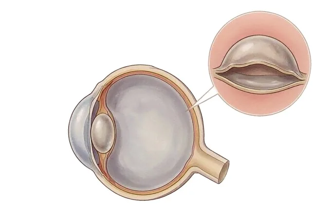
Retinal detachment is not a disease that develops quietly or a measurement that drifts out of range. It is a mechanical event: the retina, the thin sheet of light-sensitive cells at the back of the eye, comes away from the tissue that feeds it, and those cells begin to lose their supply within hours. The retina has no blood vessels of its own, which is why the separation matters so much and why it is timed rather than monitored. In most cases the sequence begins with a small tear or hole in the retina, usually in the far periphery where the retina is thinnest. Liquid gel from the middle of the eye seeps through the tear and lifts the retina off the wall, and once a sheet of retina is floating rather than attached, the region it covers stops working. Patients seldom describe this as pain, because there is none. They describe a strip of lightning at the edge of the vision, then a sudden shower of new floaters, then a shadow like the edge of a curtain that comes across the field and does not go away. The reason those three signs are treated as an emergency is the macula. The macula is the small central patch of retina that reads and recognises faces, and while it is still attached the repair can restore the situation completely. Once it has lifted, the photoreceptors in it have been starved like the rest, and some of what they carried does not come back even when the operation succeeds in putting the retina back. That is the honest framing of the condition, and it is also the practical one: this is a diagnosis that rewards speed more than almost anything else in ophthalmology, and the decision that costs sight is usually the decision to wait for an appointment.
-
The warning sequence is fixed
flashes, then a shower of new floaters, then a shadow moving across the field from the edge inwards
-
The macula sets the clock
while the centre is still attached the repair can restore the situation; once it has lifted, some sight does not return
-
An anatomical success is not a visual one
surgery puts the retina back and seals the break; it does not revive photoreceptors that have been starved
-
48 hrs
from complete records to a written assessment plan
What retinal detachment is, and why the clock matters
Retinal detachment is the separation of the retina from the tissue it sits on. The retina is a sheet of light-sensitive cells at the back of the eye, thinner than a page and with no blood vessels of its own: it is nourished entirely by the layer beneath it. When the two come apart that supply stops, and the photoreceptors begin to fail within hours rather than over months.
That single fact puts detachment in a different category from most eye disease. It is not a level to be adjusted or a process to be watched over years; it is a physical separation with a clock attached, and what the clock measures is whether the central retina is still in place.
- The cause is usually a hole in the retina. In the great majority of cases a tear or hole has opened, and liquefied vitreous gel has passed through it and lifted the retina off the wall of the eye. Because the problem is mechanical it can be closed mechanically — laser or freezing around a tear while the retina is still attached, and surgery once it is not.
- The centre of the retina is the part that matters most. The macula, the small patch that does the reading and the face recognition, keeps its function best if it is still attached when the repair is done. Once it has lifted, its photoreceptors have been starved as well, and some of what they carried does not come back even when the operation succeeds.
- None of it hurts. There is no pain, no redness and no discharge, and what is being lost is at the edge of the field, where it is easy to explain away as tiredness or a dirty lens. That is why the most useful thing a patient can do is recognise the pattern and act on it the same day.
What the operation can and cannot do. Surgery has one aim: to bring the retina back against the wall of the eye and to seal the break so that fluid can no longer get underneath it. That is a mechanical task and it is usually achievable. What no operation does is revive photoreceptors that were deprived for too long. This is why the visual outcome of a detachment is decided less by the repair than by how quickly it was carried out — and, above all, by whether the macula had already come off before it was.
The forms retinal detachment takes
Detachment is classified by what is lifting the retina, because that decides whether there is a break to seal and how the repair is planned. Three forms account for almost all cases, and one further finding sits just before them.
| What it is called | What is happening | How it behaves |
|---|---|---|
| Rhegmatogenous detachment | A tear or hole has opened in the retina, and liquefied vitreous has passed through it and lifted the retina off the wall | The commonest form by a wide margin. Treated by sealing the break — laser or freezing while the retina is attached, surgery once it has lifted |
| Tractional detachment | A fibrous membrane on the surface of the retina contracts and pulls it away; there is no tear | Seen where diabetes or another condition has grown such membranes. Often progresses more slowly, and may be watched until it threatens the centre |
| Exudative detachment | Fluid collects under the retina because of inflammation, a tumour or a vascular problem, with no break anywhere | There is nothing to seal. Treatment is directed at the underlying cause, and the retina settles as that is brought under control |
| Retinal break without detachment | A tear or hole is present, but no fluid has yet passed beneath the retina | The stage at which the problem can be closed in the clinic with laser or freezing, before any sight is lost at all |
Why the distinction is made before anything else. A retina lifted by traction has no hole to close, and a retina lifted by fluid from inflammation has nothing mechanical to correct at all. Attempting the wrong repair does not simply fail; it can make the situation worse. Establishing which of these is happening is therefore the first step of every assessment, and it is done by examining the retina itself rather than by inferring from the symptoms, which overlap heavily between the forms.
The finding that matters most is often made before a detachment exists. A tear with no fluid beneath it produces flashes and floaters and nothing else, and it can be sealed with laser in the clinic the same day. This is why patients with new and persistent flashes, or with a sudden increase in floaters, are examined urgently even when the vision is still perfect — not because a detachment is expected, but because that is the point at which it can be prevented.
What makes a detachment more or less urgent
Once a detachment is confirmed, a short list of findings determines how quickly it must be dealt with and what the patient can realistically expect. All of them are established in the same examination.
| Feature | Why it changes the plan |
|---|---|
| Whether the macula is attached or detached | The single most important determinant of what the patient will see afterwards. While the macula is on, the repair is urgent but the centre still works; once it is off, each further day affects the visual result |
| How much of the retina has lifted | A small peripheral detachment in one quadrant may be sealed with laser or supported with a localised procedure; one involving several quadrants or approaching the centre generally needs a full vitreoretinal operation |
| How long the symptoms have been present | A day and a month are different problems. A recent detachment is more likely to settle back into position and to stay there after repair |
| The state of the other eye | If the fellow eye is sound it protects the patient functionally while the affected one is repaired; if it has a break, a thinned patch or a previous detachment it is itself high-risk, and is assessed and often treated as well |
What the word urgent means here, precisely. A detachment that threatens the centre is generally operated on within days rather than hours, because the eye has to be examined properly and the right operation chosen, and that preparation is not the delay that harms. What harms is waiting weeks to report a curtain because it did not hurt.
How a detachment is assessed
The assessment answers three questions in order, and each step is chosen to make the next one precise rather than to accumulate tests.
| Step | The question it answers |
|---|---|
| History and visual acuity | What was seen, and when did it start? The sequence of flashes, floaters and shadow, together with the acuity in each eye, tells the examiner how advanced this is and whether the centre is still working |
| Examination of the retina with the pupil dilated | Where is the break, how many are there, and how much retina has lifted? Indirect examination with gentle indentation reaches the far periphery, where most breaks lie and where a direct view is of little use |
| Assessment of the macula | Is the centre attached or under fluid? Optical coherence tomography answers this precisely, and it is the finding that shapes the timing rather than a matter of impression |
| Ultrasound where the view is blocked | If blood or a dense lens hides the retina, B-scan ultrasound shows whether it is detached, where the break is likely to be, and what the operation should be — the surgical plan does not wait for the view to clear |
Why the sequence is fixed. Each answer changes the next question. Whether the macula is on or off decides the timescale; how much retina has lifted decides the operation; the position of the breaks decides where the tamponade has to press and what position the patient will have to keep afterwards. An assessment that skipped to imaging without examining the periphery would miss the finding that matters most.
Why patients travel to China for retinal detachment care
Detachment is a condition in which speed and completeness of assessment are the outcome, and what an international pathway offers is that both can happen within a single visit rather than in sequence over weeks.
The retina is examined and the answer is given the same day. Dilated examination with indentation, macular imaging and ultrasound where it is needed are all available in one facility, so a patient who arrives with a curtain in the vision leaves with a diagnosis, a plan and a date rather than a referral.
The operation can be scheduled without a waiting period. Where surgery is required, the assessment, the decision and the operation can be arranged within the same visit, which matters because in this condition the interval between diagnosis and repair is part of the treatment rather than administration around it.
Both eyes are assessed together. The fellow eye is examined at the first visit, because it shares the same anatomy and the same risks, and where a tear or a thinned patch is found it is sealed preventively in the same period instead of being left to declare itself.
The follow-up plan is written for someone going home. The schedule of retinal checks, what the vision should be doing week by week, the warning signs that require immediate attention and the point at which the eye is considered stable are provided in English; the international desk coordinates with the patient, with any insurer or assistance company, and with the treating team at home.










