


Osteomyelitis & Bone Tuberculosis Overview
Osteomyelitis & Bone Tuberculosis Overview
This page is about infection that has reached bone itself, and about why that is a different problem from infection in the soft tissues around it. Bone is rigid, so inflammation inside it raises pressure, squeezes the vessels that run through it, and leaves behind a fragment that has no blood supply. Blood supply is the only route by which antibiotics and immune cells arrive, so that fragment keeps the infection alive and keeps treatment away from it at the same time. Everything in this guide follows from that one fact: it is why an operation is part of treating this disease rather than something chosen instead of drugs, and it is why the organism has to be identified before treatment begins.
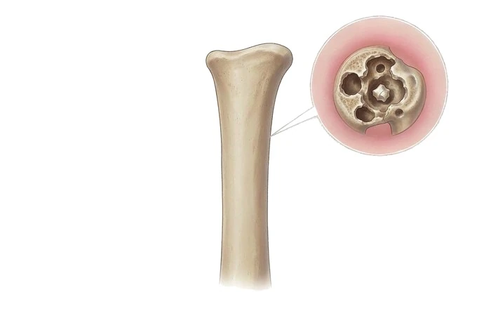
-
Dead bone is the reason it persists, not just a sign of it
a fragment without a blood supply cannot be reached by drugs or by immune cells, so it has to be removed rather than suppressed
-
Identifying the organism comes first, and before treatment
cultures taken after antibiotics have started usually tell you nothing, and the choice and length of treatment depend on the answer
-
Bone tuberculosis is a different disease, not a variant
it develops over months, produces no red hot limb, favours the spine, and is treated with a long combined regimen for which surgery has defined indications
-
A sinus that drains is not the problem, and closing it is not the cure
the opening is where the infection escapes; what has to change is what lies beneath it
What infection in bone is, and why it behaves the way it does
Infection in the soft tissues is a familiar problem: the body opens the area up, the immune system and the drugs both arrive, and it usually resolves. Bone changes the arithmetic, because bone cannot swell. Surrounded by a rigid wall, inflammation inside bone has nowhere to expand, so the pressure rises and the small vessels running through the bone are compressed. Once they close, the bone in that territory loses its supply and dies, and the body walls the dead piece off with new bone — which is a healing response that also completes the isolation.
- The dead fragment is the disease. Bone that has lost its blood supply has no immune cells working in it and no route by which a drug can reach it. It is not a scar left behind by an infection that has finished; it is a surface on which organisms survive, slowly, out of reach. This is why the illness can be quiet for months and then flare in exactly the same place, and why treating it with drugs alone so often ends in a pause rather than an end.
- Blood supply decides both the infection and the repair. The same fact explains the other half of the problem: living tissue is needed to heal, and if the tissue around the bone is poorly supplied — because of diabetes, arterial disease, scarring or previous surgery — neither a drug nor a reconstructed segment will take. This is why the assessment of the blood supply to a limb is not a preliminary to treatment; it is part of deciding what treatment is possible.
- The organism has to be identified, and the sample has to come before treatment. Bone infection is treated for a long time, and treatment given on a guess is both long and blind. A specimen taken from the surface of a sinus or an ulcer grows whatever is colonising the skin, so the specimen that matters is bone or deep tissue — taken at a procedure, or through a guided biopsy where it is safe — and it has to be obtained before the first dose, because cultures taken afterwards frequently fail to recover what was there.
- Three things have to happen, and none of them works alone. The dead bone and any infected metalwork have to be removed; the space that is left has to be covered or rebuilt with tissue that has a blood supply of its own; and the identified organism has to be treated for as long as that organism and that site require. Removing the dead bone without covering the defect leaves a surface that will not heal. Covering the defect without removing what is dead buries the problem. Getting the organism right without solving the mechanical problem produces a remission that lasts until the next flare.
The forms it takes, and why each one is managed differently
These presentations share a mechanism and differ in almost everything else: what starts them, how quickly they develop, which bone they choose, and how much of the treatment is an operation. The table below sets out the forms that account for most of the disease, and what each one commits you to.
| Form | What it looks like in practice | What this changes about treatment |
|---|---|---|
| Acute infection arriving through the bloodstream | Organisms enter the blood and settle where the flow is slowest and the supply richest: near the end of a long bone in a child, or in a vertebral body in an adult. Fever and an unwillingness to use the limb develop over a short period | A time-critical illness. The priority is to relieve pressure inside the bone and to start effective treatment quickly; a specimen is taken first wherever it can be, because the whole subsequent course depends on knowing the organism |
| Subacute infection presenting as a localised pocket | A slower, less dramatic version in which the body contains the infection inside a cavity within the bone, with weeks of local pain and little general upset | Often found on imaging done for pain that has not settled. The problem is the cavity and its contents rather than the whole bone, so the amount of bone that has to be dealt with is smaller and more precisely defined |
| Chronic infection after a fracture or an operation | An open fracture, previous surgery or metalwork that has been in place for years, followed by pain that never fully settles and often by a sinus that closes and reopens | Managed as a mechanical as well as an infective problem: the infected metalwork and the dead bone go together, the skeletal stability that the metalwork was providing has to be restored another way, and soft tissue cover becomes a central part of the plan |
| Infection beneath a foot ulcer | An ulcer on a foot whose sensation or circulation is impaired, in which the infection has reached bone. A probe that reaches bone, or persistent signs in an ulcer that is not healing, raises the question | Blood supply and pressure have to be dealt with alongside the infection, since neither drugs nor surgery will hold while the foot is poorly supplied and still being loaded through the same spot |
| Infection around an implant or a replaced joint | Pain in a replaced joint or around metalwork that does not follow the expected course, sometimes with loosening on imaging, occasionally years after the operation | Metal has no blood supply of its own, so the surfaces of an implant are exactly where organisms can persist. The strategy depends on whether the implant can be kept, exchanged in one stage or exchanged in two, which is decided by the organism, the state of the bone and the soft tissue envelope |
| Tuberculosis of bone and joint | A slow granulomatous infection that most often affects the spine, destroying disc and bone over months, with night pain, night sweats and weight loss rather than a hot swollen limb | Treated primarily with a long combined regimen directed at the organism. Surgery has defined indications rather than a routine place, and specimens have to be sent specifically for mycobacterial culture and for histology, which is what most often makes the diagnosis |
What the assessment has to establish, and what each finding commits you to
Severity in bone infection is not a grading of how ill someone feels. It is a statement about which of the problems below are present, because each one adds a commitment that cannot be met by drugs alone. The table is best read as a list of questions the assessment has to answer.
| Finding | What it means | What it commits you to |
|---|---|---|
| Infection confined to soft tissue and to the surface of the bone | Bone marrow is not involved, which is the commonest and most favourable situation, and one that imaging can usually separate from true bone involvement | A shorter course and no operation for the bone itself, provided the diagnosis is secure. The trap is treating a soft tissue infection as if bone were involved, and the reverse |
| Marrow involvement with bone that is still alive | The infection is within the bone but no fragment has yet lost its supply, which is the situation in most early cases and in many spinal infections | Effective treatment started promptly, with or without relieving pressure surgically depending on the site and the age of the patient. The aim is to prevent the step below from happening |
| Dead bone present | There is a fragment with no blood supply, sometimes visible on imaging as a dense piece within a cavity, sometimes apparent only at operation | Removal. Nothing given by any route will sterilise a piece of bone that has no circulation, so this finding is the one that turns the case from a medical problem into an operative one |
| A defect in the soft tissue envelope, or an open tract | The bone is exposed or connected to the outside through a passage, and the tissue around it has been damaged by the infection, by injury or by previous operations | Coverage with tissue that has its own blood supply, as part of the same plan as clearing the bone. Free or local tissue transfer is a solution to the dead space, not an addition to the operation |
| Instability of the bone or the spine | The infection has removed enough bone that the segment no longer holds, which causes pain with movement and prevents healing at the site | Stabilisation, either at the time of clearing the infection or in a planned second stage. Where metalwork is already present and infected, the stability it provided still has to be replaced |
| Pressure on the spinal cord or nerves | Infection, a collection of pus or a collapsed segment is narrowing the canal, with new weakness, numbness or difficulty walking | Urgent decompression. This is the one finding in the whole disease that overrides every other consideration about sequencing, and it is why new neurological symptoms in someone with a known spinal infection are treated as an emergency |
How bone infection is confirmed and followed
The investigations fall into two groups with different jobs. One group establishes what is happening now — whether bone is involved, how far it extends, whether any of it is dead. The other establishes what is causing it, which can only be answered by a specimen, and which decides the drugs and the length of treatment. Confirming the diagnosis is rarely a single test, and the sequence matters more than any individual result.
| Investigation | What it answers | What it does not answer |
|---|---|---|
| Blood tests for the inflammatory response | Whether there is an inflammatory process under way, how severe it is, and — over time — whether treatment is working | Whether bone is involved. Normal values do not exclude a bone infection, and raised values do not prove one, which is why they are followed rather than used as the diagnosis |
| Plain radiographs | Whether there is destruction, thickening or a visible fragment of dead bone, and whether an old fracture or metalwork is in the picture | Anything early. An acute bone infection usually looks normal on a plain film for the first days to weeks, so a normal film is not a reason to stop looking |
| MRI | The extent of marrow involvement, any collection of pus, any tract running to the skin, the disc space and the spinal canal, and whether the soft tissue around the bone is involved | Nothing directly, but it cannot reliably separate dead bone from living bone, and it cannot name the organism, so it defines the problem rather than the treatment |
| CT | A precise account of dead bone, the integrity of the cortex and of any fragment, the state of the spinal canal, and the geometry needed for planning an operation | Whether tissue is infected rather than simply damaged, so it is read alongside MRI rather than instead of it |
| A bone or deep tissue specimen | Which organism is responsible and what it is sensitive to, and in suspected tuberculosis whether there is granulomatous inflammation and a mycobacterial growth | Nothing at the time it is taken, because a culture takes days to weeks. This is exactly why it is taken early and before treatment, and why treatment may have to be started on a provisional basis while waiting |
Why patients travel to China for bone infection treatment
Bone infection is a problem with several specialties in it at once, and that is what most patients are actually travelling for. The assessment has to bring together imaging, microbiology, plastic and reconstructive surgery, orthopaedics and, in tuberculosis, an infectious disease and public health perspective, and the plan is only as good as the coordination between them.
- A plan made by the specialties together, before the first operation. Whether the bone has to be cleared, what is going to cover the defect, whether the skeleton needs stabilising and what organism is being treated are decisions that interact. Making them in one conversation, with the imaging and the specimens in front of everyone, is what avoids the commonest failure of this disease: a sequence of small operations that each solve one problem and leave the others alone.
- Diagnostic access within a working visit. Cross-sectional imaging, a guided biopsy where it is safe, and a laboratory able to handle both routine and slow-growing organisms can be arranged across a few days rather than across several separate referrals, which matters because the treatment cannot sensibly start before the specimens are taken.
- Reconstructive capacity where the defect is the problem. The part of this disease that is hardest to arrange elsewhere is not the removal of dead bone but what happens in the space left afterwards. Tissue transfer with its own blood supply, and later reconstruction of a segment of bone that has been lost, are the difference between a wound that closes and a limb that works.
- A single place for a long treatment, including tuberculosis. Treatment of bone tuberculosis runs for months, and the regimen has to be supervised, adjusted and completed. Being able to start, monitor and finish it in one clinical system, with the imaging and the laboratory records that go with it, removes the discontinuity that most often causes a regimen to be abandoned part-way.
- International patient services around the visit. Interpretation and accompaniment, help with visas and scheduling, an estimate of the cost of the plan in writing before it begins, direct billing with more than 30 international insurers, and follow-up by video or by written opinion once the patient has gone home. Cost sits at roughly 30% to 50% of the equivalent in Europe or the United States, and records sent in full receive a written opinion within about 48 hours.










