


Neuromyelitis Optica (NMO) Overview
Neuromyelitis Optica (NMO) Overview
Neuromyelitis optica spectrum disorder is the condition most often mistaken for multiple sclerosis, and mistaking it is expensive in a way few diagnostic errors are — because several of the medicines that treat multiple sclerosis make this one worse. The target here is not the myelin but the astrocyte , and specifically the aquaporin-4 water channel on its surface; in most people the condition is named by an antibody drawn from blood rather than by a lumbar puncture. The two signatures are the optic nerve and the spinal cord: an optic neuritis that is severe, often bilateral or rapidly sequential, and recovers less well than it does in multiple sclerosis; and a myelitis that runs lengthwise along the cord and leaves a sensory level, weakness and a bladder that has changed. A third presentation belongs in the same breath because it is the one most often missed for weeks: intractable vomiting, retching and hiccups with no explanation found in the abdomen. And the decisive fact about the prognosis is where it is made: disability here is made of attacks, not of silence — so the entire treatment question is preventing the next one, and getting this one treated within hours rather than days.
-
48 hours
written opinion once complete records arrive
-
Blood, not fluid
the antibody that names this is drawn from a vein
-
Attacks
where the disability is made — not silence between them
-
124+
countries and regions our international patients come from
What neuromyelitis optica is
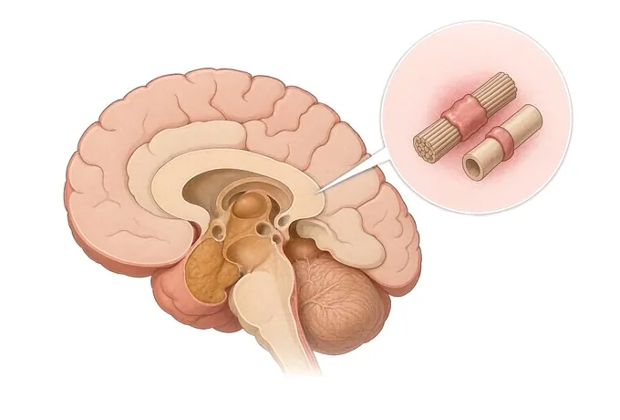
The name is old and it is not a good one, because it describes the two places the condition most often shows itself — the optic nerve and the spinal cord — rather than what it is. What it is, is an autoimmune disease in which the target is the astrocyte: a support cell of the central nervous system, carrying on its surface a water channel called aquaporin-4. An antibody binds to that channel, complement is activated, the astrocyte is injured, and the nerve fibres that depended on it are damaged secondarily. That is a different sequence from what happens in multiple sclerosis, where the immune attack is directed at the myelin and at the axon beneath it.
The practical consequence of that difference is not academic. It is the reason the two conditions are named by different tests, followed by different imaging, and treated with different medicines — and it is the reason several of the agents used in multiple sclerosis, including some of the most effective ones, are associated with worse outcomes in this condition rather than better ones.
What the condition looks like, then, is a small number of recognisable attacks rather than a slow decline. An optic neuritis that takes vision severely and gives back less than the optic neuritis of multiple sclerosis does. A myelitis that runs along the cord rather than across it, leaving a level below which sensation is altered and the limbs and the bladder behave differently. And, less often and later recognised, an attack on the area postrema at the base of the brainstem which presents as vomiting and hiccups and is investigated for weeks in the wrong place.
Two sentences carry the rest of this guide. The first: the disability in this condition is made of attacks rather than of silence, which is unlike multiple sclerosis and is the reason the treatment question here is almost entirely about preventing the next one. The second: the attack that is happening now is the one that will be lived with, which is why treatment within hours rather than days is not a nicety.
The three names, and why they are not interchangeable
Three labels are in circulation and they are regularly used as though they were one. They are not, and what separates them is what the blood shows.
| Name | What the blood shows | What the distinction changes |
|---|---|---|
| AQP4-IgG-positive NMOSD | Aquaporin-4 antibody detected by cell-based assay on serum | The diagnosis is made, and maintenance immunosuppression is the treatment — not the standard multiple sclerosis agents |
| MOG antibody disease (MOGAD) | MOG antibody detected, with the aquaporin-4 antibody negative | A different condition with a different course: attacks are often steroid-responsive, and the tendency to relapse is lower than in AQP4-positive disease |
| Seronegative NMOSD | Neither antibody detected, at least on the assays and at the time it was drawn | A stricter combination of clinical and imaging features is required, and the antibody may be worth repeating during a relapse |
One practical point about that last row, because it is the one that costs people years. A negative antibody result is not the same as no antibody. The assay matters — cell-based assays perform differently from older methods — the timing matters, and the result can be negative while someone is on immunosuppression. Where the clinical picture fits and the test was negative, it is worth repeating during a relapse rather than closing the question.
What makes an attack urgent
In multiple sclerosis, a relapse is usually a matter of days to arrive and weeks to resolve, and it is rarely a reason to be in an emergency department tonight. In this condition the arithmetic is different, because an attack is where the disability is made. What follows is what changes the timescale, and what each one warrants.
| Feature | What it warrants |
|---|---|
| Visual loss that is severe from the outset, or that involves both eyes within days | Assessment now, and the antibody drawn before any treatment is started. Bilateral and sequential optic neuritis is characteristic here in a way it is not in multiple sclerosis |
| A sensory level on the trunk, weakness in the legs, and a bladder that has stopped working properly | Imaging of the cord now, with contrast. This is the shape of a longitudinally extensive myelitis, and the hours matter |
| Vomiting, retching or hiccups lasting days with no gastrointestinal cause found | Not a gastroenterology problem once the abdomen has been looked at. This is the area postrema syndrome, and it belongs in this pathway |
| Weakness or sensory change that is at its worst within seconds or minutes | The shape of a stroke rather than of an inflammatory attack. Emergency assessment now, and the question of which condition this is asked afterwards |
| A deterioration with fever, or someone becoming hard to rouse | Infection or another acute condition until shown otherwise. Both worsen this condition and both are missed for it |
| Symptoms that come on with heat and resolve within the hour | Not an attack. The same damage conducting worse at a higher temperature, which is a management problem rather than a reason for more immunosuppression |
| A slow increase in stiffness and slowing over a year, with no attack anyone can point to | Not the usual shape of this condition. Where it happens, the structural causes of a compressed cord are excluded before anything else is assumed |
The error to avoid is the reverse of this table as well: an attack being watched for a few days to see whether it resolves on its own. In a condition where the disability is made of attacks, waiting is not neutral — it is the single most available way to make the outcome worse.
How the diagnosis is assembled
The assessment is not a single test that comes back positive. It is a clinical pattern, an imaging finding, and — in most people — an antibody in the blood. Alongside them sits a second job that is done far less often than it should be: excluding the conditions that look like this one, multiple sclerosis most of all.
| Item | What it establishes | What it does not do |
|---|---|---|
| Aquaporin-4 antibody on serum, cell-based assay | Names the condition in most people who have it, and separates it from multiple sclerosis | Not a test done on spinal fluid, and not a result that is fixed for ever. It can be negative on one assay and positive on another, or negative while immunosuppressed |
| MOG antibody on serum | Names a different condition where the aquaporin-4 antibody is negative, and changes the expected course | Not a test that means the same thing in every clinical setting. A transient positivity in a child after a single episode is read differently from a persistent one |
| MRI of the cord, with contrast | A longitudinally extensive lesion — three or more vertebral segments — which is characteristic here and unusual in multiple sclerosis | Not seen at all where only the brain is imaged. This is the commonest technical omission in the workup of this condition |
| MRI of the brain | Excludes a great deal, and in this condition is frequently normal or shows change that does not satisfy multiple sclerosis criteria | Not the study that makes the diagnosis. A normal brain scan in someone with a cord lesion and positive antibodies is entirely compatible with this condition |
| MRI of the orbits with contrast | Involvement of the optic nerve, and how far along it the signal runs | Not always abnormal, and an old optic neuritis can leave the nerve looking unremarkable years later |
| Spinal fluid | Excludes infection and other inflammation; the cell count and protein are often raised during an attack | Oligoclonal bands are absent in most people with this condition. Their presence should prompt the multiple sclerosis question again rather than settle this one |
| Blood tests for the mimics | The autoimmune and infectious conditions that produce the same picture, and the thyroid and lupus antibodies that travel with this one | Not a substitute for the antibody test itself. A file full of negative autoimmune screens with no AQP4 result is a file in which the decisive test was not sent |
| The history, in order | What happened first, how fast, whether it recovered and how completely — which is often where the second attack, and therefore the dissemination, is hiding | Not a formality. An episode six years ago that was never written down is regularly the most valuable item in the file |
Why patients travel to China for a neuromyelitis optica assessment
Most of what families are looking for here is not a new treatment. It is an answer to a question that was never asked: whether the aquaporin-4 and MOG antibodies were ever tested, and whether the treatment that has been running is the right one for the disease that is actually there. That question is answered from records more often than not, which is why the first step is a written opinion rather than a journey.
Where the person does need to be here, what an assessment adds is the assembly: neurology, imaging, rehabilitation and neuropsychology contributing to one plan rather than four letters, with the cord and the orbits imaged as a matter of course rather than on request. The programme is supported by 3.0T magnetic resonance imaging, dual-source computed tomography, positron emission tomography, three-dimensional digital subtraction angiography, long-term video electroencephalography, the full spinal fluid set, and evoked potentials — and by antibody testing arranged as part of the workup rather than as an afterthought.
What should not be assumed is that a particular medicine is available here. Where a medicine recommended is not carried out within this pathway, that is named at the outset, before any payment is discussed — not at the end of a workup. Where the honest answer is that the plan already running is the right one and nothing here would change it, that is said as plainly as the alternative, and it is not converted into an itinerary.










