


Nasopharyngeal Carcinoma Overview
Nasopharyngeal Carcinoma Overview
Nasopharyngeal carcinoma sits behind the nose, where it can grow for months without being seen, and it is unusually sensitive to radiation. That combination makes it a disease where the sequence of treatment matters more than the speed of an operation.
-
60–80%
of patients first notice a painless lump in the neck
-
EBV
Epstein-Barr virus — implicated in almost all non-keratinizing tumours
-
IMRT
intensity-modulated radiotherapy, the standard technique
-
48 hrs
from complete records to a personalized MDT plan
What nasopharyngeal carcinoma is
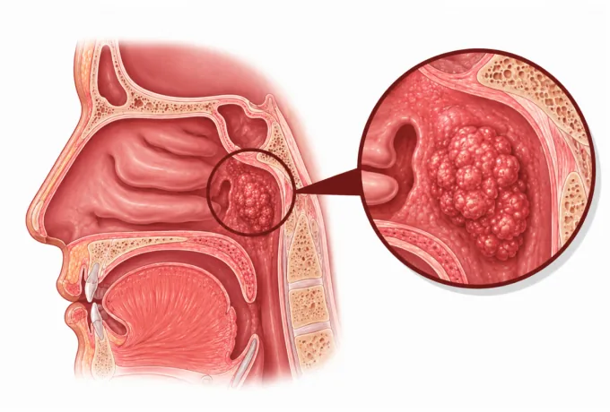
The nasopharynx is the space behind the nose, above the soft palate, where the nasal airway turns down into the throat. It is small, it opens into the eustachian tubes that ventilate the middle ear, and it sits directly beneath the skull base. Nasopharyngeal carcinoma arises from the epithelial lining of this space — most often in a recess on the side wall called the fossa of Rosenmüller, which is why the tumour can grow for a long time without blocking the nose or causing anything the patient would notice.
Two features make this cancer different from most head and neck cancers. The first is its relationship with Epstein-Barr virus: in the non-keratinizing types that dominate in southern China and Southeast Asia, viral DNA and viral gene products are found in essentially every tumour cell. The second is its radiosensitivity. Nasopharyngeal carcinoma responds to radiation to a degree that squamous cancers elsewhere in the head and neck do not, which is why radiotherapy — not surgery — is the treatment that cures it, and why an operation on the primary tumour is rarely the right first move.
Where you grew up therefore matters more for this cancer than for almost any other. It is uncommon in most of Europe and North America, and among the most common cancers in southern China, Southeast Asia and North Africa. That geographic pattern follows a mixture of viral exposure, childhood diet and inherited susceptibility — and it is also why treatment experience with the disease is concentrated in the same regions.
The main types
The World Health Organization divides nasopharyngeal carcinoma into three main groups, and the group is not academic — it predicts how the tumour behaves and how it responds to treatment:
- Keratinizing squamous cell carcinoma — the type more common in Europe, North America and older patients, linked to tobacco and alcohol rather than to the virus, and the least responsive to radiotherapy.
- Non-keratinizing carcinoma, differentiated — the commoner non-keratinizing pattern in endemic regions, strongly EBV-associated and markedly radiosensitive.
- Non-keratinizing carcinoma, undifferentiated — the classic endemic type, almost entirely EBV-driven, and the most radiosensitive of all; it also has the highest rate of spread to neck nodes, which is why a neck lump is so often the presenting sign.
- Basaloid squamous cell carcinoma — a rarer variant with a distinctive microscopic appearance and a less favourable outlook than the non-keratinizing types.
- Salivary gland-type carcinoma — a small group of tumours of the nasopharynx arising from minor salivary tissue, treated more like salivary gland cancer than like nasopharyngeal carcinoma.
The practical difference is the third group. An undifferentiated non-keratinizing tumour can melt away under radiotherapy in a way that a keratinizing tumour will not, and it tends to spread to the lymph nodes early rather than to invade locally. That is the reason the treatment plan for nasopharyngeal carcinoma is built around the radiotherapy target and the neck, rather than around the question of how much tissue to remove.
What each stage means
Staging is done with MRI of the nasopharynx and skull base rather than CT, because the questions that decide the stage are soft-tissue questions: has the tumour reached the parapharyngeal space, has it eroded the skull base, has it grown towards the cranial nerves. The AJCC system then groups the tumour (T), the nodes (N) and distant spread (M) into four stages.
| Stage | In plain terms | How it is usually approached |
|---|---|---|
| I | Tumour confined to the nasopharynx, with no involved nodes and no distant spread | Radiotherapy alone — intensity-modulated radiotherapy to the nasopharynx and the neck |
| II | Tumour extending into the parapharyngeal space, or nodes in the upper neck on one or both sides | Radiotherapy, with concurrent chemotherapy in many cases |
| III | Tumour eroding the skull base or reaching the paranasal sinuses, or nodes on both sides of the neck | Concurrent chemoradiotherapy; induction chemotherapy before it in selected cases |
| IVA | Tumour reaching inside the skull, the cranial nerves, the orbit or the lower throat, or extensive neck nodes | Induction chemotherapy followed by concurrent chemoradiotherapy |
| IVB | Distant spread — most often to bone, lung or liver | Systemic therapy, including a PD-1 inhibitor; radiotherapy to sites causing symptoms |
| Recurrence | Disease returning at the primary site or in the neck after radiotherapy | Decided by where and when it returned — surgery, re-irradiation, or systemic treatment |
Two details in this table carry most of the prognostic weight. Skull-base erosion is the boundary between a tumour that can be covered comfortably by a radiotherapy plan and one that needs the plan built around the brain and the optic nerves. And neck node involvement is present in the majority of patients at diagnosis, which is why the neck is treated as part of the target rather than as a separate problem.
Epstein-Barr virus and the markers that guide treatment
Nasopharyngeal carcinoma has fewer actionable molecular targets than lung or breast cancer, and one marker that matters more than any of them: Epstein-Barr virus DNA in plasma.
| Marker | What it is | How it changes management |
|---|---|---|
| Plasma EBV DNA | Viral DNA released by tumour cells and measurable in a blood sample | Used before diagnosis to screen high-risk people, at diagnosis as a staging and prognostic marker, and after treatment to detect recurrence months before imaging |
| EBV in tumour tissue | Viral RNA and protein detectable in the biopsy | Confirms the diagnosis in a non-keratinizing tumour and separates it from a keratinizing cancer |
| PD-L1 | An immune checkpoint protein reported from the biopsy | Supports the use of a PD-1 inhibitor in recurrent or metastatic disease, and is being tested in earlier stages |
| Plasma EBV DNA clearance | The rate at which viral DNA falls during treatment | A slow fall or a rise after treatment is a signal to look harder for residual or recurrent disease |
The reason EBV DNA matters so much is that it is measurable from a blood test. In an endemic region it can be used to screen people with no symptoms; during treatment it tracks how well the tumour is responding; and after treatment a rising level is a more sensitive early warning than a scan, because it reflects tumour burden rather than anatomy. If a biopsy was taken elsewhere and the tumour type is unclear, testing the tissue for EBV is usually the first thing to request.
Why patients come to China for nasopharyngeal carcinoma care
Nasopharyngeal carcinoma is unusual in that the highest incidence in the world is in southern China — Guangdong, Guangxi and Fujian — which means the greatest concentration of treatment experience is in the same place. A centre that treats this disease every week plans the radiotherapy target differently from one that sees it a few times a year, and the difference shows up in both tumour control and in the side effects the patient lives with afterwards.
- Intensity-modulated radiotherapy at volume — the standard technique for this disease, used to give a curative dose to the nasopharynx and the neck while keeping the dose to the salivary glands, brainstem, optic nerves and temporal lobes within tolerance.
- Plasma EBV DNA in routine practice — used for screening, for staging, for monitoring the response during treatment and for surveillance afterwards, which changes how recurrence is detected.
- Concurrent and induction chemotherapy — cisplatin given with radiotherapy, and gemcitabine-based induction regimens before it, planned by medical oncology alongside the radiation oncologist.
- PD-1 inhibitors — several agents in this class were developed and approved in China and are available for recurrent or metastatic disease.
- Surgery in its proper place — neck dissection for residual nodes after radiotherapy, and endoscopic nasopharyngectomy for selected local recurrence, rather than an operation on the primary tumour at diagnosis.
Two practical points follow from how the disease behaves. Because radiotherapy takes weeks, the plan should be written before travel rather than during it — send complete records and the written assessment is returned within 48 hours of the file being complete, including a view on whether induction chemotherapy is indicated. And because the side effects of treatment are cumulative and partly reversible with early management, follow-up matters as much as the treatment itself.










