


Multiple Sclerosis (MS) Overview
Multiple Sclerosis (MS) Overview
Multiple sclerosis is an immune-mediated disease of the central nervous system, and the two words that carry the diagnosis are disseminated and in time . The damage has to appear in more than one region — the optic nerve, the brainstem, the spinal cord, the white matter around the ventricles — and it has to have happened on more than one occasion, or be present on one scan as both old and new. Neither is a formality; both are the thing that separates this condition from the much commoner finding of a few white matter lesions on a scan done for a headache. Where the lesion sits decides what the symptom is: one nerve produces painful loss of vision, the brainstem produces double vision and imbalance, the cord produces a limb and a bladder. And every episode leaves two different things behind — the relapse, which is the event, and the residue, which is what stays . Disability in this condition is made mostly of residue rather than of episodes. This guide follows the order people need it in: what each course looks like, what the symptoms actually cost, how the diagnosis is assembled and what it takes to exclude, what changes the long-term course and what only treats the week, and what follow-up has to contain — including the half that does not appear on any scan.
-
48 hours
written opinion once complete records arrive
-
One team
neurology, imaging, rehabilitation and neuropsychology together
-
Dissemination
in space and in time — what the name actually requires
-
124+
countries and regions our international patients come from
What multiple sclerosis is
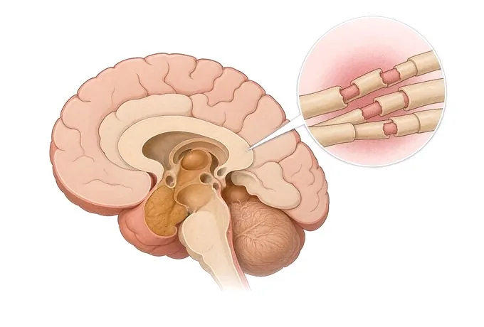
Nerve fibres in the brain and the spinal cord are wrapped in myelin, and the myelin is what lets a signal travel quickly along them. In multiple sclerosis the immune system damages that wrapping and, to a varying degree, the fibre underneath it. The damage happens in patches, the patches are scattered rather than in one place, and over time they leave scars — which is where the name comes from.
What follows from that is the whole of the diagnostic question. Because the damage is scattered, the disease has to be shown to be scattered: lesions in more than one characteristic region of the central nervous system. Because the damage happens in episodes, it has to be shown to have happened more than once: a new lesion appearing on a later scan, or old and new lesions sitting together on one scan, or — in the right clinical setting — the pattern in the spinal fluid that goes with this disease. These two requirements are called dissemination in space and dissemination in time, and they are not paperwork. They are what separates this condition from the far commoner situation of someone being told they have MS because a scan showed a few bright spots.
Where the patch sits decides what the person experiences, which is why two people with the same disease can look nothing alike. A lesion on the optic nerve produces blurred or lost vision in one eye, usually with pain on moving it. A lesion in the brainstem produces double vision, or a room that will not stop moving. A lesion in the spinal cord produces numbness or weakness in the limbs and a bladder that has started behaving differently. Lesions in the cerebral white matter, which are the ones most often seen on a scan, may produce nothing a person can name at all.
And every episode leaves two different things behind. The relapse is the event — days to develop, weeks to improve, and often improved substantially. The residue is what is still there afterwards: a foot that catches, an eye that never quite got its colour back, a bladder that warns later than it used to. Disability in this condition is made mostly of residue accumulating, not of episodes happening, and that is why the treatment page separates what changes the long-term course from what treats an episode. What each of those looks like day to day is set out on the symptoms page.
The courses, and what each one decides
Which course a person has decides what the treatment is aiming at, and it is worth being precise because the names are used loosely. What separates them is not severity but shape: whether the disease arrives in episodes, whether it accumulates quietly between them, and whether it has been accumulating from the beginning.
| Course | What it looks like | What the distinction changes |
|---|---|---|
| Relapsing-remitting | Episodes that develop over days and improve over weeks, with periods between them in which nothing new appears | The commonest course at the outset, and the one in which disease-modifying therapy has the clearest role |
| Secondary progressive | A relapsing course that, after years, is joined by a slow accumulation of disability — with or without further relapses | The question becomes what is still inflammatory and what is residue; and whether the current therapy still fits |
| Primary progressive | Accumulation from the outset, without the early relapses that make the pattern obvious | Later to be named, and most often first mistaken for something structural — a cord being compressed, or ageing |
| Clinically isolated syndrome | One episode — an optic neuritis, a myelitis, a brainstem episode — in someone with no previous history | Not yet this disease. What is decided here is whether dissemination can be shown, and whether to start treatment before it can |
| Radiologically isolated syndrome | Lesions with the shape of this disease found on a scan done for another reason, in someone with no symptoms at all | The honest answer is not to treat. It is to say what has been found, what it does not yet mean, and what to watch for |
Two rows carry most of the consequence in practice, and they pull in opposite directions. Clinically isolated syndrome is the one in which something is genuinely at stake: a first episode is the moment at which a decision about long-term treatment may be made, and it is the moment most often lost — the episode resolves, the person is discharged, and the question of what else was on the scan is never answered. Radiologically isolated syndrome is the one in which the right action is restraint: a scan with suggestive lesions in someone with no symptoms is not a diagnosis, and starting treatment on it commits a person to years of monitoring for a disease they may never have. Naming both matters more here than in most conditions, because the error runs in both directions.
What changes the timescale
Two clocks run here, and confusing them is how the urgent thing is allowed to wait and the ordinary thing is turned into an emergency. The first is the clock of a relapse: days to arrive, weeks to resolve, and rarely a reason to be in an emergency department tonight. The second is the clock of everything that is not a relapse but looks like one — a stroke, a cord being compressed, an infection — which is measured in minutes to hours. What follows separates them, with the action attached to each.
| Feature | What it looks like | What it warrants |
|---|---|---|
| Maximal at the onset | Weakness, loss of speech or loss of vision that is at its worst within seconds or minutes of starting | That is the shape of a stroke, not of a demyelinating episode. Emergency assessment now, and the MS question asked later |
| A new deficit lasting more than a day | Numbness, weakness, double vision or visual loss that developed over a day or two and has not gone | Assessment now rather than at the next appointment, and a relapse considered. Not an emergency, but not a wait either |
| A sensory level, leg weakness and bladder involvement | A band of altered sensation across the trunk, legs that will not do what they are told, and urine that will not come or will not wait | Imaging of the cord now. This is also the shape that neuromyelitis optica takes, and the two are not managed the same way |
| Visual loss with pain on eye movement | Blurred or lost vision in one eye, aching behind it when the eye moves, colour that looks washed out | Optic neuritis, and the question is what else is on the scan — because a first episode is a decision point rather than a footnote |
| Fever, or a falling level of consciousness | An acute deterioration with a temperature, or a person who is becoming harder to rouse | Not a relapse. This is infection or another acute inflammatory condition until shown otherwise, and it belongs in a different pathway |
| Worse in the heat, better when cooled down | Vision or strength that goes off in a hot shower, on a hot day, or with a fever, and returns within the hour | Not a relapse, and not a new lesion. Conduction failing at temperature — which is a management problem rather than a treatment decision |
| Stiffness and walking that have been declining for a year | Legs that are stiff and slow, a gait that has narrowed, and no episode anyone can point to | Primary progressive until shown otherwise — and the structural causes of a compressed cord excluded before that name is used |
The error to avoid is the reverse of this table: a person with a sudden maximal deficit being told it is probably a relapse because they have MS, or a person with a genuine relapse being told to wait for their next scheduled scan. Both happen regularly, and both are prevented by asking one question before anything else — did this reach its worst in minutes, or in days? They are repeated on the symptoms page, because that is where they get used.
How the diagnosis is assembled
The assessment is not a single test that comes back positive. It is a set of findings that, together, either demonstrate dissemination in space and in time or do not — and alongside them, a set of tests whose real job is to exclude the conditions that look like this one. Both halves are necessary, and the second is the one most often left undone.
| Step | What it establishes | What it does not do |
|---|---|---|
| The history, taken as episodes | What happened, in what order, how long it took to arrive, and whether it fully went — the dissemination in time is often here rather than on any scan | Not a formality. A symptom that came and went six years ago and was never written down is the single most valuable item on this page |
| The neurological examination | Which pathways are involved, including the ones the person has not noticed — an absent abdominal reflex, a pale disc, a slightly ataxic heel-shin | Not a substitute for imaging, and not normal just because the person walks into the room unaided |
| MRI of the brain, with contrast | Lesions in the characteristic regions, and whether any of them is enhancing — which means active now rather than at some point | Not a count that tracks how a person feels. Lesion burden on a scan correlates poorly with disability |
| MRI of the spinal cord | Cord lesions, which are the commonest way dissemination in space is completed and the commonest thing omitted from a brain-only scan | Not optional where the symptoms point below the neck. A brain scan alone will miss the lesion that answers the question |
| Spinal fluid | Oligoclonal bands present in the fluid but not in the blood — supportive of this disease — and the exclusion of infection and other inflammation | Not required in every case, and not diagnostic alone. A normal fluid does not exclude the disease |
| Blood tests | The mimics: aquaporin-4 and MOG antibodies, and the inflammatory, infectious and nutritional causes that produce the same picture | Not a screening panel to be ticked. Each is requested because it changes management, and the antibody tests change it most |
| Evoked potentials | Slowing in a pathway the person has no symptoms in — which can supply the second region when the examination and imaging do not | Not a first-line test where imaging has already answered the question, and not a diagnosis on its own |
| Follow-up imaging | A new lesion, or a new enhancing lesion — the second occasion, which is what time requires | Not a scan done because a year has passed. It is done because a question is open, and it is compared against the previous one |
What a finished assessment ends in is a written judgement of four things: whether dissemination has been demonstrated, and on what; which alternatives have been excluded and on what grounds, naming the antibody tests specifically; what would change the answer, including whether a repeat scan or a repeat of the fluid is the right next step; and what the follow-up has to contain — the intervals at which imaging is repeated, what symptoms are asked about at each visit, and what would prompt a change in treatment. The full assessment, including what it costs and how long it takes, is set out on the diagnosis page.
Why patients travel to China for a multiple sclerosis assessment
Patients come for four reasons. The first is a scan that was reported as suggestive and never followed up: white matter lesions found on imaging done for a headache, a sentence in a letter that says demyelination cannot be excluded, and no one who has said what happens next. The second is a diagnosis that was made without the alternatives being excluded — most often without the aquaporin-4 and MOG antibody tests, which is a different disease with a different treatment and a different prognosis. The third is a first episode that resolved and was discharged, where the question of what else was on the scan was never answered. And the fourth is the half that no scan shows: fatigue, heat sensitivity, slowed thinking and bladder function that have quietly reorganised a working life and have never been formally assessed.
What the pathway looks like. Neurology and imaging plan together, with rehabilitation and neuropsychology inside the same programme. 3.0T MRI, dual-source CT, PET-CT, 3D DSA and long-term video EEG sit within it, as do the full cerebrospinal fluid set — chemistry, cell differential, oligoclonal bands — and the antibody tests that separate this condition from the neuromyelitis optica spectrum. A written opinion is returned within 48 hours of complete records arriving. Fully treated costs typically run at 30–50% of comparable care in Europe or the United States, more than 30 commercial insurers are billed directly, and our international patients come from 124 countries and regions.
What must be said about the medicines and the procedures. Corticosteroids for a relapse, disease-modifying therapy, the medicines used for spasticity, pain, bladder function and fatigue, plasma exchange where a severe relapse has not responded, and lumbar puncture itself are each discussed as they sit in this pathway: what they are used for, what monitoring they require, and what their own risks are. No medicine is presented as one this hospital routinely holds, and no therapy is presented as one that ends the matter. Any medicine or procedure recommended that is not carried out within this pathway is named at the outset, before any payment is discussed — not at the end of a workup.
What a records-only opinion can and cannot do. It can say whether the imaging demonstrates dissemination in space and in time, whether the spinal fluid was interpreted in the context of what was actually sent, which antibody tests were done and which were not, whether a follow-up scan was ever compared against the first, and what the follow-up has so far omitted. It cannot examine the person, and examination is where several of these distinctions are made: the optic discs, the reflexes, the gait, the sensory level and the visual acuity cannot be assessed from a file. We say so rather than implying otherwise.
What to send first. The imaging with its report — the images rather than a description of them, and both the brain and the cord where both were done. Alongside them: any spinal fluid result with its date, the list of what has been given and for how long, and a short account in your own words of each episode in the order it happened, including what recovered and what did not.










