


Multiple Myeloma Overview
Multiple Myeloma Overview
Multiple myeloma is a cancer of plasma cells, the cells in the bone marrow that make antibodies. Because every malignant cell descends from one original cell, the antibody they produce is a single identical protein rather than the usual mixture of thousands of different ones. That protein is what makes myeloma so measurable: a blood or urine test can detect it, quantify it, and follow it over years of treatment. It is also what damages the kidney in some patients, because light chains filtered by the kidney are toxic to the tubules when produced in large quantities.
-
Protein
a single abnormal antibody found in the blood or urine — the reason myeloma can be measured rather than only seen
-
CRAB
calcium, kidneys, anaemia and bone: the four ways the disease causes damage, and the findings that separate active myeloma from a form needing no treatment
-
Marrow
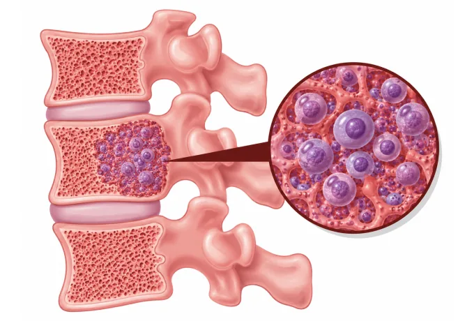
and the bone around it: plasma cells crowd out normal blood production and activate the cells that break bone down
-
48 hrs
from complete records to a personalized MDT plan
What multiple myeloma is
A plasma cell is a specialised white cell whose whole purpose is to make one antibody, chosen after an immune challenge and produced in quantity. Myeloma begins when one such cell acquires the changes that let it multiply without being needed, and its descendants inherit the same antibody. The plasma cells themselves stay in the marrow, so they do not form a lump that can be felt; the protein they make, however, travels in the blood and passes into the urine, and that is what a laboratory can detect. The consequence is that myeloma announces itself through a test result far more often than through a physical finding.
Two mechanisms cause the harm. The first is crowding: plasma cells occupy marrow space and suppress the production of red cells, white cells and platelets, so anaemia is the commonest abnormality found at diagnosis, and infections are a frequent problem. The second is signalling: myeloma cells activate the osteoclasts that resorb bone and inhibit the osteoblasts that form it, producing areas of bone loss that weaken the skeleton rather than adding to it. Those lesions release calcium into the blood, and that raised calcium, together with the light chains passing through the kidney, accounts for much of the damage the disease does to organs.
That is why the four classic features are grouped under the acronym CRAB — calcium, renal impairment, anaemia and bone lesions — and why the diagnosis is framed around them. Myeloma is not staged by how far cells have travelled, as a solid tumour is, but by how much damage has occurred and by a small number of laboratory values that predict how the disease will behave. A patient is not treated because plasma cells are present; a patient is treated when they are present in sufficient quantity and something on that list has occurred.
The spectrum, from monitoring to treatment
Myeloma is unusual in that its early forms are not cancer in any practical sense, yet they are the reason a diagnosis is often made before symptoms start. The conditions are separated by two measurements — how much monoclonal protein is present, and what proportion of the marrow is occupied by plasma cells — together with whether any damage has occurred:
- Monoclonal gammopathy of undetermined significance — a small monoclonal protein with fewer than 10% plasma cells in the marrow and no damage. It is common in older adults, it is not treated, and it is followed at intervals because a small proportion of cases progress over years rather than months.
- Smouldering multiple myeloma — a larger protein or a larger plasma cell proportion, but still no damage. It too is monitored rather than treated, because treating it before damage appears has not been shown to help; the monitoring interval is set by how much protein is present and by the chromosome findings.
- Active multiple myeloma — the disease that requires treatment, defined by the presence of plasma cells together with at least one of the defining events described below. This is what is usually meant by the word myeloma in a treatment plan.
- Solitary plasmacytoma — a single collection of plasma cells in bone or in soft tissue, without plasma cells in the marrow or a monoclonal protein elsewhere. It is treated with radiotherapy to that site, and followed because a proportion of patients later develop myeloma.
- Light chain amyloidosis — a related condition in which light chains produced by the plasma cells deposit in organs such as the kidney, heart or nerves and interfere with their function. It can occur with myeloma or on its own, and because the organs involved limit what treatment can be given, it changes the plan substantially.
- Variants that change the testing rather than the treatment — some patients produce only light chains, which are found by the free light chain assay rather than by electrophoresis, and a small number produce no detectable protein at all, in whom the marrow and imaging carry the diagnosis alone.
Staging and what separates active disease
Two different questions are answered by two different assessments, and confusing them is a common source of misunderstanding. One is whether the disease needs treating at all; the other is how advanced it is once it does. The first is answered by the defining events, the second by the staging systems:
| Category | In plain terms | How it is used |
|---|---|---|
| The CRAB features | A raised calcium level, impaired kidney function, anaemia, or bone lesions that are visible on imaging | Any one of these, together with plasma cells, makes the disease active and means treatment starts |
| The three laboratory thresholds | Plasma cells occupying 60% or more of the marrow, a free light chain ratio of 100 or more, or more than one area of bone involvement on MRI that is not yet visible on plain imaging | These indicate disease that is about to cause damage, and treatment is offered even though no organ damage has occurred yet |
| International Staging System | Stage I to III, calculated from two blood tests: beta-2 microglobulin and albumin | Records how advanced the disease is when treatment begins, and is used to set the intensity of a plan |
| Revised International Staging System | The same stages, with two additions: the lactate dehydrogenase level, and whether any of three specific chromosome findings is present | Separates the patients within each stage whose disease is likely to behave more aggressively, and who may need more intensive treatment from the outset |
| Smouldering myeloma | A raised protein or a raised plasma cell proportion, with none of the damage or threshold findings above | Observed rather than treated, with the monitoring interval chosen from the protein level and the chromosome results |
| Myeloma-related organ involvement | Deposition of light chains in organs, most often the kidney, heart or nerves, which is a separate diagnosis that may accompany myeloma | Changes which treatments can be used, because the organs involved determine what the patient can tolerate |
The point worth drawing out is that the threshold matters more than the diagnosis. Two patients with identical plasma cell proportions in the marrow may be told very different things — one that the disease will be watched and the other that treatment starts next week — because of a calcium value or an imaging finding. This is not an inconsistency but a consequence of how the disease damages the body: it is the damage, or the near certainty of it, that justifies treatment, because the treatments carry their own cost in side effects.
It also means that a plan should state the stage and the reason for treatment rather than the diagnosis alone. Asking which features put the disease into the active category, what the beta-2 microglobulin and albumin were, and whether the chromosome findings raised the stage are all reasonable questions at the outset, because each of them changes what is recommended.
The markers that guide treatment
Myeloma has no single defining mutation, and that shapes how its genetics are used. The chromosome findings do not make the diagnosis, which rests on the plasma cells and the damage; instead they sort patients into those whose disease behaves predictably and those whose disease behaves aggressively, and they occasionally point to a drug that would otherwise not be considered:
| Marker | What it is | How it changes management |
|---|---|---|
| Rearrangements involving chromosome 14 | The antibody-producing genes on chromosome 14 are placed next to an activating gene, which drives the plasma cell to multiply — the commonest type of change in myeloma | Whether the partner gene is one of the two high-risk partners decides whether the disease is classed as higher risk and whether the initial treatment is intensified |
| Loss of chromosome 17p | The loss of a region where a gene that normally restrains damaged cells sits | Associated with a less durable response to standard treatment, which is a reason to favour continuous combination treatment and to plan monitoring closely |
| Extra copies of chromosome 1q | Additional copies of part of chromosome 1, found in a substantial proportion of patients | Associated with a greater chance of relapse, and taken into account when deciding the sequence and duration of treatment |
| The chromosome 11;14 rearrangement | A rearrangement that is common in myeloma and is also characteristic of a related plasma cell condition | Identifies patients whose disease may respond to a drug that acts on a pathway this rearrangement makes the cells dependent on |
Two further measurements follow the disease rather than describing it. The monoclonal protein level in the blood and the free light chain ratio are measured at every assessment, and the depth of their fall is how a response is judged — a complete response means the protein can no longer be detected by the standard tests. When the protein disappears entirely from the blood and urine, testing the marrow for residual plasma cells with a sensitive method gives a fuller answer, and that result is increasingly used to decide how long treatment should continue.
The practical instruction is to ask for the chromosome and molecular report itself rather than the conclusion, because the stage and the risk group are calculated from it, and because some findings indicate a specific drug. It is worth asking, in writing, which of the reported changes affected the plan — both what was recommended and what was left out.
Why patients come to China for myeloma care
Myeloma care is a long relationship rather than a single treatment, and it depends on capabilities that are either present as a coherent service or are not: a laboratory that measures the protein and the light chains reliably and repeatedly, a transplant programme, access to the newer drug classes, cell therapy, and the supportive care that keeps the kidneys and the skeleton safe through years of treatment.
- Reliable monitoring of the protein — electrophoresis, immunofixation and free light chain assays available in-house, since these numbers are the measure of every treatment decision and cannot be reconstructed retrospectively.
- Current combination regimens — treatment built from three drug classes, with an antibody directed at CD38 added in the first line, chosen by whether a transplant is planned rather than by one regimen applied to everyone.
- Autologous stem cell transplant — a programme with apheresis, a stem cell laboratory and transplant wards, offered to patients whose disease has responded and whose fitness allows it.
- Cell therapy and bispecific antibodies — treatments directed at BCMA, for disease that has returned after several lines, which are the options of greatest interest to patients who have already been treated elsewhere.
- Kidney and bone protection as part of the plan — correction of calcium, avoidance of the drugs and contrast agents that harm the kidney, bone-strengthening medication, radiotherapy or cement injection for painful lesions, and pain control.
- Imaging that shows what plain X-rays miss — whole-body low-dose CT or MRI at diagnosis, because the number and site of bone lesions change the treatment and because plain films under-report them.
Because myeloma is treated continuously, the practical arrangements matter more than in most cancers. Patients who live elsewhere typically have the initial assessment and the intensive part of treatment in China, then continue maintenance closer to home under a written schedule, returning for defined reviews. The records that make that possible are specific: the protein electrophoresis and immunofixation reports with the values, the free light chain ratio with both values and the units, the marrow report with the plasma cell proportion and the chromosome findings, the imaging report, and a treatment history with dates and the response at each assessment. Send those, and the written assessment is returned within 48 hours of the file being complete.










