


Moyamoya Disease Overview
Moyamoya Disease Overview
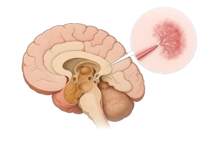
Moyamoya disease is a progressive condition of the blood vessels supplying the brain. The terminal portions of the internal carotid arteries — the last stretch before they divide at the base of the skull — narrow over years and eventually close, and the same happens to the origins of the arteries that arise from them. As the main channels close, the brain grows its own alternative supply: a fine network of small vessels at the base of the brain. On a catheter angiogram that network has a hazy, indistinct look, and the name of the disease comes from the Japanese word for it.
-
National
Stroke Center construction unit, and a national GCP base
-
3D DSA
Alongside 3.0T MRI, dual-source CT, PET-CT and 24-hour video EEG
-
48 hours
Written second-opinion report after complete records arrive
-
124+
Countries and regions our international patients come from
What the narrowing is, and where it happens
The narrowing is at a specific place, and naming it is the first step towards reading a report. The internal carotid artery enters the skull and ends by dividing into the anterior and middle cerebral arteries. In moyamoya disease it is the terminal segment of the internal carotid that narrows, together with the first part of the two arteries it gives rise to. The process is symmetrical in the sense that matters for diagnosis: it affects both sides. Where it affects one side only, the condition is described as probable or unilateral moyamoya, and is followed rather than assumed to be the same illness.
The vessel wall changes in a recognisable way. The inner layer thickens, with a proliferation of cells and a distorted internal elastic lamina, while the muscular layer thins. The artery does not become blocked by a clot travelling from elsewhere, and it is not the same process as the fatty plaque of atherosclerosis — the patients are often children,
The collateral network is the part that gives the disease its name, and it is worth understanding what it is and what it is not. These vessels are the brain's attempt to supply itself: perforating branches at the base of the brain enlarge and multiply, and transdural and transcranial channels open as well. They carry real blood and they are why many patients function at all. But they are small vessels operating at pressure they were not designed for, and they are the source of much of the bleeding in adults. A network that looks reassuring on a scan is a sign that the main vessels have already closed, not a sign that the circulation is secure.
One thing should be said early, because it changes how everything else is read. Moyamoya disease is progressive. A patient diagnosed at five with mild narrowing will not have the same vessels at fifteen, and a patient whose scan looks stable today may still be losing reserve. This is the single most important difference between this condition and the other structural cerebrovascular conditions in this group, and it is the reason the question in moyamoya is rarely "should we wait and see".
Ischaemic or haemorrhagic, child or adult, disease or syndrome
Moyamoya is sorted in three ways at once, and each one changes the plan.
Ischaemic or haemorrhagic. A patient whose events have all been transient ischaemic attacks or strokes is described as having the ischaemic form, and a patient who has bled has the haemorrhagic form. The distinction matters because the treatments and the urgencies differ: bleeding is an event to be survived and then prevented from recurring, while ischaemia is a process to be outpaced. Children overwhelmingly present with ischaemia. Adults present with either.
Child or adult. There are two peaks in age at presentation: one in childhood, around five years of age, and one in adulthood, in the fourth decade. They are not the same illness seen at different ages. The childhood form progresses faster, causes repeated transient attacks, and can affect development and cognition when it is not treated. The adult form progresses more slowly and carries the risk of bleeding, which is the presentation in a substantial minority and the reason the randomised trial of surgery in adults enrolled patients who had already bled.
Moyamoya disease or moyamoya syndrome. The same angiographic pattern can appear in the setting of another condition — sickle cell disease, neurofibromatosis type 1, Down syndrome, previous radiotherapy to the head, thyroid disease, and several autoimmune conditions among them. When one of these is present the term moyamoya syndrome is used, and the management is of both the vascular picture and the underlying condition.
How far it has gone, and how much reserve is left
Two separate things are being measured, and they are not the same number. One is anatomy — how much of the main vessel has closed, and what has grown to replace it. The other is physiology — whether the circulation can still increase its flow when the brain asks it to. A patient can have advanced anatomy and adequate reserve, and a patient can have modest anatomy and none. The second is what decides timing.
| Axis | How it is described |
|---|---|
| How far the narrowing has gone | Described in stages on catheter angiography, from narrowing of the terminal carotid alone through to complete occlusion of the main channels with the collateral network fully developed and, at the far end, with the collateral supply itself receding as it too fails. |
| Whether both sides are involved | Bilateral, or unilateral. Bilateral involvement is required for the diagnosis of moyamoya disease under the widely used criteria; a unilateral picture is called probable moyamoya disease and is followed with imaging, because a proportion of these patients go on to develop changes on the other side. |
| How far the collateral network has developed | Sparse, established, or receding. These vessels are what is keeping the brain perfused, and their appearance on an angiogram is read alongside the perfusion studies rather than on its own. |
| Whether an aneurysm is present | Aneurysms occur on the collateral vessels and on the perforating arteries more often in moyamoya than in the general population, and when bleeding has occurred they may be the source rather than the fragile collaterals themselves. Identifying one changes the plan directly, because the aneurysm is treated on its own account and the revascularisation is planned around it. |
| Ischaemic or haemorrhagic presentation | Whether the events so far have been transient attacks and strokes, or a bleed. This is the axis that most changes what surgery is being asked to do: in the ischaemic form the aim is to prevent the next stroke and to protect development, while in the haemorrhagic form the aim is to reduce the chance of bleeding again, |
| How much reserve is left | Whether the vessels can still dilate when demand increases, measured with perfusion imaging before and after a challenge agent such as acetazolamide. A circulation with exhausted reserve is one in which the brain is already running at capacity: it cannot respond to a fever, to dehydration or to a drop in blood pressure. This is the measurement that most often moves a decision from observation to surgery. |
| Stage of treatment, and time since it | Untreated, recently operated, or revascularised and established. After indirect surgery the new vessels take weeks to months to grow in, so the months after an operation are a period of transition rather than of protection, and the antiplatelet and hydration precautions continue through it. After direct surgery the flow is immediate, and the concern in the first days is the opposite one: too much flow. |
Read together, these axes do not answer "how severe is this". They answer which of three situations this is: a patient with exhausted reserve and symptoms, in whom surgery is being discussed because the next event is anticipated rather than merely possible; a patient found on a scan with no symptoms and preserved reserve, in whom the question is how closely to follow and whether the other side is involved; or a patient who has bled, in whom the work is to establish the source and then to reduce the chance of it happening again.
How moyamoya is demonstrated
The sequence begins with the non-invasive studies and proceeds to angiography when the question is no longer whether but how far — and, in most patients, to a perfusion study, because the anatomy alone does not say whether the brain is coping.
| Investigation | What it settles |
|---|---|
| MRI and MR angiography | Whether the terminal carotid arteries are narrowed or closed, whether the collateral network at the base of the brain is present, and whether there is evidence of injury already sustained — an infarct, or changes the patient never had symptoms from. In many centres this pair of studies is sufficient to establish the diagnosis, |
| Digital subtraction angiography | The reference study, and the one on which the extent and the stage are defined. It shows the vessels filling in sequence, which is how the collateral network is distinguished from the main circulation, and it shows the external carotid supply that a bypass would use. It is performed by the team that would carry out the operation, because the images are the plan. |
| Perfusion imaging, with an acetazolamide challenge | Whether the circulation can still increase flow on demand. Acetazolamide dilates the small vessels; where the reserve is intact, flow rises after the challenge, and where it is exhausted, it does not, and may fall in territories that are stealing from each other. This is the study that turns an anatomical finding into a decision, and it is the one most often missing from a workup done elsewhere. |
| CT of the head, and CT perfusion | Whether bleeding has occurred, and where the blood lies — in the brain tissue, in the ventricles, or in the space around the brain — because the site points towards the source. CT perfusion gives a second view of reserve where acetazolamide challenge is not available, and it is the study used in the emergency setting. |
| EEG, in a child | A characteristic pattern in which slowing appears during and after hyperventilation and then resolves — a finding that supports the diagnosis and that can be obtained without sedation. |
A report worth having therefore answers six things: whether both sides are involved, how far the narrowing has gone, how well the collateral network is developed, whether there is an aneurysm, whether the disease is ischaemic or haemorrhagic in this patient, and how much reserve is left — the last of which is the one that decides whether the discussion is about surgery now or about follow-up.
Why patients travel to China for moyamoya surgery
A patient who is having transient attacks, or who has just bled, needs care where they are, and no one should travel during an event. Travel becomes reasonable between events, and in three situations: when the diagnosis has been made and the question is whether revascularisation is indicated and by which technique; when a patient has been told to wait and wants a second reading of the perfusion studies, which are the part most often absent from a workup; and when the need is rehabilitation and cognitive support after a stroke or a bleed.
Our neurology and neurosurgery services are organised as a standardised stroke centre built to national stroke-centre standards, and are a National Stroke Center construction unit and national GCP base. Cerebrovascular cases are planned jointly by neurology, neurosurgery and rehabilitation rather than handed from one department to the next, and interventional work — moyamoya disease, arteriovenous malformations, aneurysms and arterial stenosis — is carried out on 3D DSA, alongside 3.0T MRI, dual-source CT, PET-CT and 24-hour video EEG.
The practical questions are answered in the same way as for any other condition we treat. Records reviewed by a neurovascular specialist receive a written opinion within about 48 hours of complete records arriving, and the opinion states what the vessels show, how much reserve remains if it has been measured, whether revascularisation is indicated and by which technique, and what remains uncertain. Complete treatment in China typically runs at 30–50% of comparable care in Europe or the United States. We work with more than 30 commercial insurers for direct billing and have received patients from 124 countries and regions.










