


Meniscus Tear Overview
Meniscus Tear Overview
Each knee has two menisci: wedge-shaped rings of fibrocartilage that spread load across the joint, help it stay stable and help it glide. When one of them tears, the first thing that matters is not how big the tear is but what shape it has and where it sits, because only the outer rim has a blood supply and only a tear with a blood supply has any prospect of healing. The second thing is whether the tear is actually the source of the complaint: locking, catching and giving way behave quite differently from an ache, and scans of knees that do not hurt show meniscal change often enough that a finding on its own proves very little. The third is what else is in the joint, because osteoarthritis and a ligament injury change both the plan and the target. Removing tissue and repairing it are two different operations with two different aims, and how much of the meniscus is kept is the part that matters for the joint decades later.
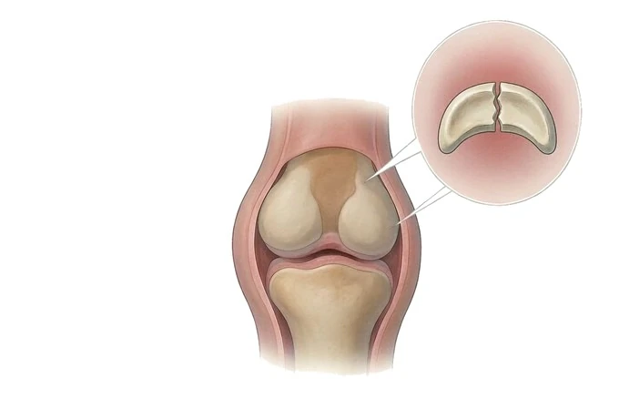
-
The pattern decides whether it can heal
a tear in the vascular rim is a different problem from one in the centre
-
Where it sits matters as much as how it looks
the outer rim bleeds; the inner margin does not
-
Not every tear is the source of the pain
tears are common on the scans of knees that do not hurt
-
Removing is not the same as repairing
what is kept matters to the joint decades later
What the meniscus is, and what a tear actually means
The menisci are two wedge-shaped rings of fibrocartilage sitting between the thigh bone and the shin bone, one on the inner side of the knee and one on the outer. They spread load, they help the joint stay stable, and they help it move smoothly. A tear in one of them is one of the commonest things that happens to a knee, and it is also one of the most over-treated, because what the word means varies enormously.
- The meniscus is not padding. It converts a rounded bone end meeting a flat surface into something closer to a socket, spreading the load over a larger area. Taking tissue away is not removing something dispensable; it changes how the joint carries force.
- Blood supply is not uniform, and this is the fact that organises everything. The outer rim has vessels reaching it. The inner margin does not. A tear in the first has some prospect of healing; the same tear in the second has none, because nothing can deliver the cells and the signal that healing requires.
- The shape of the tear matters as much as its size. A tear running lengthways in the vascular rim can be brought together and held. A tear cutting across the rings destroys the hoop that resists load. A tear with a fragment that flips into the joint blocks movement. Same organ, three different problems.
- Finding a tear and finding the cause of the pain are two different tasks. Meniscal change is common on the scans of knees that do not hurt, and becomes more common with age. A scan is useful when it agrees with what the person described and with what was found on examination, and it is misleading when it is read on its own.
What this means practically is that the useful questions are not "is there a tear". They are what pattern it has, where it sits, whether the complaint is mechanical or an ache, and what else is happening in the joint. Those four answers, rather than the scan alone, are what a plan is built from.
The patterns a tear can take, and why the shape is named first
Meniscal tears are named by pattern and by position, and the name is not descriptive habit: it is what determines whether the tear can be repaired, whether it blocks the joint, and whether it has taken away the meniscus's ability to do its job.
| Pattern | What it looks like | What it implies |
|---|---|---|
| Longitudinal (vertical) | Runs lengthways, parallel to the rim, usually in the vascular outer zone | The pattern most often suitable for repair, particularly in a younger knee |
| Bucket-handle | A longitudinal tear whose inner fragment flips into the middle of the joint | Frequently causes true locking and a block to straightening; usually needs dealing with |
| Radial | Cuts across the meniscus from the inner edge outwards, dividing the hoop | Removes the tension the meniscus depends on; often not repairable in the way a rim tear is |
| Horizontal (cleavage) | Splits the meniscus through its thickness, often running to the surface | Commonly degenerative; often managed without surgery unless it produces mechanical symptoms |
| Flap or parrot-beak | A piece that is partly detached and can move into the joint | Prone to catching; the movable piece is often the part addressed |
| Complex | More than one pattern at once, often in tissue that has already worn | Usually degenerative; repair is often not realistic |
| Root tear | Detachment where the meniscus anchors to the bone | Takes the meniscus out of function, so the joint behaves as though it has been removed; treated seriously rather than watched |
| Discoid meniscus | A thicker, disc-shaped meniscus present from birth, usually on the outer side | A shape variant rather than an injury; may present with clunking in a child and is evaluated rather than assumed to be a tear |
Two of these are worth holding apart in the mind, because they are the ones most often confused. A longitudinal tear in the rim and a horizontal split in worn tissue can both be called a meniscus tear and neither may be the reason the knee hurts; but the first may be worth repairing, while the second usually is not.
Two axes: whether this tear can heal, and what it is doing to the person
Severity in a meniscus tear is read on two axes that do not substitute for one another. The first is about the tissue: can this tear heal? The second is about the person: what is this knee no longer able to do?
The first axis — the biology of the tear. What determines whether a repair has anything to work with is blood supply, and blood supply follows position.
| Zone | Blood supply | What it means |
|---|---|---|
| Red–red (outer rim) | Vessels reach it from the capsule | The zone in which a repair has a biological basis |
| Red–white (middle) | Partial supply | An intermediate situation, weighed case by case |
| White–white (inner margin) | No direct supply | Healing cannot be relied on; removal of the unstable piece is more often the option |
The second axis — what the knee can no longer do. This is the axis that decides treatment, and it is not measured on the scan.
- Mechanical symptoms. True locking, in which the knee will not come straight; giving way under load; a catch that recurs in the same movement. These point at something physically in the way.
- An ache. Pain that is worse at night, worse after walking a distance, worse coming down stairs, and that settles with rest. This pattern belongs more often to the joint surface and to osteoarthritis than to the tear.
- What has been given up. Which activity has stopped, how long for, and what has already been tried. A tear that has not changed what someone can do is a different proposition from one that has ended a season.
- How long it has been going on. A knee that has been avoiding load for weeks loses muscle, and the wasting is part of what makes it feel unreliable.
Putting the two axes together is the whole planning problem. A repairable tear in a knee with mechanical symptoms is one thing. A degenerative tear in a knee with osteoarthritis and an ache is quite another, and the second is usually managed without an operation first.
What has to be established before a plan is chosen
A knee is assessed by putting three sources next to one another and checking that they agree. Where they disagree, that disagreement is the finding.
- The history, and specifically whether the complaint is mechanical or an ache. Locking, catching and giving way are asked about directly, because they are volunteered less often than pain is.
- Examination for joint-line tenderness. Tenderness along the strip where the meniscus sits is the finding most consistently associated with a tear, and it takes seconds to establish.
- Whether the knee can straighten fully. A block to the last few degrees of extension suggests something mechanically in the way, and it is more informative than most of the special tests.
- The ligaments, at the same appointment. A missed ligament injury changes what any meniscus treatment has to withstand; the two are injured together often enough that looking for one and not the other is an error.
- Weight-bearing radiographs. They do not show the meniscus. They show the joint it lives in, including whether osteoarthritis is present, and that often determines the plan more than the tear does.
- Magnetic resonance imaging, interpreted against the symptoms. It shows the pattern and the position. It is misleading when read on its own, because meniscal change is common in knees that do not hurt and becomes more common with age.
- The joint as a whole. Alignment, the muscles around it, and what the person needs the knee to do. A tear is treated inside a knee, not in isolation.
What a complete assessment has to produce is not a description of the tear but four answers: what pattern it is and where it sits; whether the symptoms fit the tear or fit the joint instead; what else is going on, particularly osteoarthritis and the ligaments; and what has already been tried and for how long. With those four in front of them, most patients can see for themselves why a plan is being proposed.
Why this matters when choosing where to be treated. The commonest failure in meniscus care is not technical. It is a scan being treated as a diagnosis, followed by an operation on a finding that was never causing the symptom. Next in this guide: the diagnosis page sets out how the history, the examination and the scan are read against one another, and the treatment page sets out what repair and partial removal each involve.
Why patients come to China for knee surgery
Patients weighing treatment abroad are usually comparing three things: whether the assessment will be done carefully enough to know what is actually causing the symptom, what will be done to the meniscus itself, and whether the plan can continue after they go home.
- A written opinion within about 48 hours of complete records arriving. Send the history, the radiographs and any magnetic resonance imaging already done, and the team will set out what the scan shows, whether the symptoms appear to fit it, and what the reasonable options are. That opinion is also what a patient uses to decide whether travelling is worthwhile before anyone books a flight.
- Repair considered before removal. Where the tear sits in the vascular rim and the pattern allows it, bringing the edges together is discussed first, because what is kept matters to the joint decades later.
- The joint treated, not just the tear. Ligaments are examined at the same time, osteoarthritis is part of the plan rather than an aside, and alignment and muscle are addressed rather than left for someone else.
- Rehabilitation on a standardised pathway. Recovery across our network follows a standardised nine-node pathway, so the plan set out at the end of treatment is the one followed afterwards, and it travels with the patient in writing.
- Cost and insurance. Treatment in China generally costs around 30% to 50% of comparable care in Europe or North America. More than thirty international insurers are billed directly across our network, so the patient does not pay upfront and claim afterwards; whether a particular policy is one of them is confirmed from the policy details rather than assumed.
- Practical support. The international patient team assists with appointments, interpretation, visa invitation letters and local logistics, and patients from more than 124 countries and regions have been treated across our network of 19 institutions.










