


Meningioma Overview
Meningioma Overview
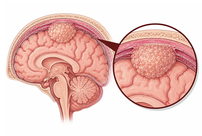
A meningioma does not begin in the brain. It begins in the meninges — the layers of tissue that wrap around the brain and the spinal cord — and it grows outwards from that covering, pushing the brain aside rather than growing within it. That single difference from a tumour of the brain tissue itself explains most of what makes a meningioma a different problem: it usually has a border that a surgeon can follow, complete removal is often achievable, and the site of the tumour determines which functions are at risk rather than its size alone.
-
Commonest
the most frequent tumour that begins inside the skull in adults
-
Outside in
it grows from the covering of the brain, pushing the brain aside rather than growing within it
-
Grade and site
what the tissue shows and where the tumour sits — the two findings that decide the plan
-
48 hrs
from complete records to a personalized MDT plan
What a meningioma is
The brain and spinal cord are wrapped in three layers of tissue called the meninges, whose outer layer lies against the inside of the skull. Between the layers are cells — the arachnoid cap cells — from which a meningioma arises. Because the tumour grows from a covering rather than from the brain itself, it displaces the brain instead of infiltrating it, and it usually carries its own blood supply and its own capsule. All of that has practical consequences.
The first consequence is that the tumour can grow considerably before it causes anything. The brain is not invaded function by function; it is compressed gradually, and the nervous system accommodates a slow change. This is why so many meningiomas are found incidentally, on a scan performed for a headache, a minor injury or an investigation of something else entirely, and why the finding of one is not automatically a reason for immediate treatment.
The second is that the site matters more than the size. A tumour over the outer surface of the brain may reach several centimetres with no more than a seizure as its first sign. A tumour the size of a grape at the base of the skull, pressing on the nerve that carries sight or on the nerves for hearing and balance, or narrowing one of the large veins that drain the brain, can cause a serious deficit while still small. Where the tumour sits decides which functions are at risk, and therefore what the treatment has to protect.
The third is that the diagnosis is usually made from the scan rather than from the tumour. The appearance of a meningioma on contrast-enhanced MRI is characteristic, so tissue is often not needed before a plan is made. What tissue does provide — and this becomes important the moment treatment is contemplated — is the grade, because grade decides the likelihood of recurrence and therefore the intensity of the treatment that is appropriate.
The grades, and where tumours occur
Meningiomas are grouped in two ways that matter clinically, and a patient will usually be told both:
- By grade. Most are grade 1: slow-growing tumours whose cells look much like normal arachnoid cells, and which are cured by complete removal in a large proportion of cases. Grade 2, or atypical, meningiomas show features that predict a greater chance of returning — a higher rate of cell division, or invasion into the brain tissue next to the tumour. Grade 3, or anaplastic, tumours are the rarest and behave most aggressively, and are treated with surgery plus radiation from the outset.
- By histological variant. The pathologist names the tumour for its appearance: meningothelial, fibrous and transitional are the commonest grade 1 types; psammomatous tumours contain the small calcified bodies seen on CT; secretory and angiomatous are further variants. A few variants carry their own implications — clear cell and chordoid tumours are more likely to return, and papillary and rhabdoid tumours are treated as higher grade.
- By site — over the surface of the brain. Convexity tumours sit on the outer surface, and parasagittal or falcine tumours lie along the midline against the large vein that runs there. Both can often be removed completely, though the midline position makes the vein the structure that limits how far removal can safely go.
- By site — at the base of the skull. Sphenoid wing, olfactory groove, tuberculum sellae, cerebellopontine angle and petroclival tumours lie against nerves and vessels. They are often the ones that cause symptoms early, and the ones where removal has to be balanced against the risk to sight, hearing, smell or the blood supply to the brain.
- By site — other locations. Tumours may arise within the ventricles, along the optic nerve sheath, or in the spinal canal, and each has its own pattern of symptoms and its own set of treatment considerations.
Grading and the extent of removal
Meningiomas are not staged in the way cancers of other organs are. Two measures do the work instead — the grade given by the pathologist, and how completely the tumour was removed:
| Category | In plain terms | How it is used |
|---|---|---|
| Grade 1 | A slow-growing tumour whose cells resemble the normal cells of the covering of the brain | Complete removal is the aim, and for small tumours that cause nothing, observation is a legitimate alternative |
| Grade 2 (atypical) | Features that predict a greater chance of the tumour returning — a higher rate of cell division, or invasion of the brain next to the tumour | Followed more closely after surgery, and radiation is considered even when removal appears complete |
| Grade 3 (anaplastic) | The rarest group, with the most aggressive behaviour | Treated with surgery and radiation together, and followed most closely of all |
| Extent of removal | How much of the tumour, and of the covering it arose from, the surgeon was able to take | The strongest predictor of whether the tumour returns, and the reason the operation is planned in detail beforehand |
| Site | The structures the tumour lies against — the large veins, the nerves of sight, smell, hearing and balance, and the arteries at the base of the brain | Determines which functions limit how far removal can go, and therefore how much tumour is likely to be left behind |
| Brain invasion | Whether the tumour has grown into the brain tissue next to it, rather than only pressing on it | A finding that raises the grade regardless of how the tumour otherwise looks, and changes the follow-up plan |
The practical point is that the grade is not known until the tissue has been examined. A tumour that looks entirely ordinary on the scan may turn out to be atypical, and the operation that appeared to be the whole of the treatment may then need to be followed by radiation. This is why the pathology report should be read for the grade and for its reason, and why a report that gives only the tumour's name without a grade is an incomplete report.
The findings that predict recurrence
Molecular testing in meningioma does not define the diagnosis the way it does in some other brain tumours, but in tumours that are not clearly grade 1 it does influence how closely the patient is watched and how ready the team is to treat again:
| Finding | What it is | How it changes management |
|---|---|---|
| NF2 alteration | A change in a gene that controls the growth of these cells, found in a substantial share of meningiomas and central to neurofibromatosis type 2 | Confirms the biology of the tumour, and in a patient with the associated condition, sets the surveillance programme for the rest of the nervous system |
| CDKN2A/B deletion and TERT promoter mutation | Two changes associated with more aggressive behaviour | Mark tumours that are likely to return sooner, and support treating them as higher grade even when the appearance alone is not decisive |
| BAP1 loss, SMARCE1 and other changes | Findings that occur in particular variants of meningioma and in some families | Point to the variant, and occasionally to an inherited predisposition that needs its own follow-up |
| AKT1, SMO, KLF4 and TRAF7 changes | Changes found in a subset of grade 1 tumours, some of which are located in particular sites | Help to classify a tumour whose appearance is not characteristic, and are the subject of research into drug treatment for tumours that recur |
Two practical conclusions follow. First, in a clearly grade 1 tumour that has been completely removed, molecular testing rarely changes anything, and its absence from a report is not a gap. Second, in a tumour that is atypical, that has been incompletely removed, or that has returned, the molecular findings are worth having, because they help decide how closely to follow the patient and how quickly to consider further treatment. Asking which situation applies to you is a reasonable question to put to the team.
Why patients come to China for meningioma care
The care of a meningioma is decided by a small number of capabilities that have to work together, and the balance between them is what the treatment consists of: a plan that is either surgical or not, an operation that is either complete or limited, and a decision about radiation that is made on the basis of both.
- Imaging that establishes what is safe to remove — contrast-enhanced MRI together with imaging of the large veins, since it is the position of the tumour against the venous sinuses and the nerves at the base of the skull that decides how far an operation can go.
- Surgery planned around the structures at risk — skull base approaches, the techniques that let the tumour be separated from the brain and from the nerves it has displaced, and preoperative embolisation for tumours with a rich blood supply.
- Removal that aims at the whole of the tumour and the involved covering — because the extent of removal is the strongest predictor of whether the tumour returns, and because leaving the involved meninges behind is what makes a recurrence likely.
- Precision radiation when surgery is not the right answer — stereotactic radiosurgery for small tumours or for the part left behind after an operation, and fractionated radiotherapy for larger tumours or those lying against structures that cannot tolerate a single high dose.
- Neuropathology that reports the grade and its reason — because grade 2 or 3 changes the follow-up and makes radiation part of the initial plan rather than something held in reserve.
- Rehabilitation and symptom control — physiotherapy, speech and language therapy, cognitive support and control of seizures, which determine how much function is recovered after an operation near critical structures.
Because the treatment of a meningioma is often a long sequence with imaging at intervals rather than a single episode, patients who live elsewhere commonly have the diagnosis reviewed and the operation or the radiation performed in China, then return home with a written schedule for the imaging that follows. The records that make this workable are the MRI images and reports with dates, the pathology report with the grade, the operative report with the extent of removal, the radiotherapy plan with dose and volumes if treatment has been given, and the current medication. Send those, and the written assessment is returned within 48 hours of the file being complete.










