


Lymphoma Overview
Lymphoma Overview
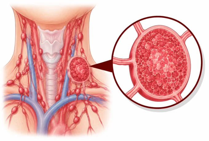
The lymphatic system is the body's drainage and surveillance network: lymph nodes connected by vessels, with the spleen, the thymus and the marrow as its larger organs, and lymphocytes circulating through all of it. Because that network is everywhere, lymphoma can begin almost anywhere — and because it begins in the immune cells themselves, its treatments are as much immunology as chemotherapy. A painless node that stays enlarged is the usual first sign; it is a finding to have examined, not a finding to wait on.
-
~90%
of lymphomas are non-Hodgkin lymphomas, and these are dozens of distinct subtypes rather than one disease
-
PET/CT
maps the whole body in one scan, because most lymphomas take up the tracer — and the same scan later confirms response
-
Painless
enlarged lymph nodes are the commonest first sign; a node that does not settle is the finding to have examined
-
48 hrs
from complete records to a personalized MDT plan
What lymphoma is
Lymphocytes are the white blood cells that recognise and remember threats. They are produced in the marrow, schooled in the thymus, and stationed in lymph nodes, the spleen and the lymphoid tissue of the gut, skin and airway. Lymphoma begins when one of those cells acquires the changes that let it divide without limit — and because lymphocytes are designed to travel, the resulting disease is often widespread at the moment it is found, which is why staging examinations the whole body rather than one region.
The distinction that matters more than any other is between Hodgkin lymphoma and non-Hodgkin lymphoma. They are separated under the microscope rather than by how they present: Hodgkin lymphoma contains Reed-Sternberg cells, spreads in an orderly way from one node group to the next, and is among the most treatable of all cancers. Non-Hodgkin lymphomas lack those cells and behave according to their subtype — some grow over days, others over years.
That difference is not academic. A high-grade lymphoma such as diffuse large B-cell lymphoma changes size week to week and is treated promptly with combination immuno-chemotherapy, because delaying it costs the chance of cure. An indolent lymphoma such as follicular lymphoma can be widespread yet slow, and treating it early has not been shown to prolong life — so the correct plan may be to watch it closely rather than to start treatment. The same finding in two patients can therefore lead to opposite advice. What determines which applies is the subtype, established by an excision biopsy of a whole lymph node. Needle sampling alone is usually insufficient, because it collapses the nodal architecture that defines the diagnosis.
The main types
Lymphoma is classified by the cell it arises from and by its behaviour. The list below covers the diagnoses most patients are given, with what distinguishes each:
- Classical Hodgkin lymphoma — defined by Reed-Sternberg cells, often presenting as painless nodes in the neck or as a mass in the chest. It spreads predictably, takes up PET tracer brightly, and is highly responsive to chemotherapy, radiotherapy and checkpoint inhibitors.
- Nodular lymphocyte-predominant Hodgkin lymphoma — a separate, slower entity that often presents as a single node and is managed differently from classical Hodgkin lymphoma.
- Diffuse large B-cell lymphoma — the commonest aggressive lymphoma, typically a rapidly enlarging mass in a node, the abdomen or elsewhere, and treated with curative intent from the outset.
- Follicular lymphoma — the commonest indolent lymphoma, often already widespread at diagnosis with nodes that wax and wane; the practical question is when treatment is worth starting.
- Marginal zone lymphomas — gastric, splenic and nodal forms, several of which arise in the setting of chronic infection or of an autoimmune disease.
- Mantle cell lymphoma — usually nodal and usually advanced at diagnosis, with a biology that keeps it returning; continuous targeted treatment has changed how it is managed.
- Burkitt lymphoma — among the most rapidly dividing of all human tumours, requiring intensive treatment and specific measures to protect the nervous system.
- Small lymphocytic lymphoma — the nodal and tissue counterpart of chronic lymphocytic leukaemia, and treated as the same disease.
- Peripheral T-cell and NK/T-cell lymphomas — a heterogeneous group including angioimmunoblastic T-cell lymphoma, the ALK-positive and ALK-negative anaplastic large-cell lymphomas, extranodal NK/T-cell lymphoma of nasal type, and cutaneous T-cell lymphoma affecting the skin.
- High-grade B-cell lymphoma with rearrangements of MYC and BCL2 or BCL6 — the so-called double-hit and triple-hit lymphomas, treated more intensively than standard diffuse large B-cell lymphoma.
Three practical consequences follow. The subtype determines whether treatment begins this week or is monitored for a period. It determines whether the regimen contains an antibody, a checkpoint inhibitor, a kinase inhibitor or cell therapy. And because subtype names are specific — follicular and diffuse are different diseases, not degrees of one — it is worth asking for the full name rather than accepting "lymphoma" as an answer.
What each stage means
Staging for lymphoma uses the Lugano classification, which is built on the older Ann Arbor system and adapted to what imaging now shows. Most lymphomas take up the tracer used in PET-CT, so a single scan can describe every involved site — which is why a stage in lymphoma describes distribution rather than tumour size:
| Category | In plain terms | How it is usually approached |
|---|---|---|
| Stage I | One lymph node region, or one site outside the nodes | Short-course systemic treatment, or radiotherapy alone for limited disease in selected subtypes |
| Stage II | Two or more node regions, all on the same side of the diaphragm | Combination immuno-chemotherapy, with radiotherapy added for some early presentations of Hodgkin lymphoma |
| Stage III | Node regions on both sides of the diaphragm | Full-course combination treatment, with a mid-treatment PET-CT used to check that the disease is responding |
| Stage IV | Involvement of marrow, liver, lung or another organ beyond the nodes | Full-course systemic treatment; transplant or cell therapy considered in selected cases |
| A and B | B symptoms — fever, drenching night sweats, weight loss — are recorded alongside the stage letter | Their presence places the disease in a higher risk group and changes how intensively it is treated |
| Risk group | The International Prognostic Index, the follicular index or the Hodgkin score combines stage with age, blood results and fitness | Selects which regimen is used, and whether treatment is intensified or maintenance is given afterwards |
Two features of that table are worth drawing out. Stage alone does not determine treatment in lymphoma the way it does in most solid tumours: a stage III indolent lymphoma may need no treatment at all, while a stage I aggressive lymphoma is treated immediately and with curative intent. That is why the risk group sits alongside the stage — it converts the same stage number into different recommendations depending on age, fitness and blood results, and why a plan that quotes a stage without a subtype and a risk group is not yet a plan.
The second feature is that staging is repeated, not done once. Because most lymphomas are FDG-avid, a scan partway through treatment can show whether the disease has stopped taking up the tracer — the single strongest early indicator of how the disease is responding — and the result may lead to continuing, changing or shortening the regimen. When a scan is not informative, as in some indolent and low-grade subtypes, staging and response are judged on CT and on the clinical picture instead.
The markers that guide treatment
Lymphoma is defined by the proteins its cells display, so testing is not an optional extra added late — it is how the diagnosis is made. A short list of targets then determines which treatments are open:
| Marker | What it is | How it changes management |
|---|---|---|
| CD20 | A protein on the surface of nearly all B cells and of most B-cell lymphomas | The target of rituximab and obinutuzumab, which are built into standard first-line treatment for B-cell lymphoma |
| CD30 | A protein on Reed-Sternberg cells in Hodgkin lymphoma and on some T-cell lymphomas | The target of brentuximab vedotin, used in Hodgkin lymphoma and in anaplastic large-cell lymphoma |
| CD19 | A protein present on B cells from an early stage of their development | The target of CAR T-cell therapy and of bispecific antibodies for relapsed or refractory B-cell lymphoma |
| MYC with BCL2 or BCL6 rearrangement | Two or three gene rearrangements occurring together in the same tumour | Identifies high-grade B-cell lymphoma, treated more intensively and with specific measures to protect the nervous system |
Beyond those four, several other results change decisions in specific situations. In Hodgkin lymphoma, the PD-1 ligands are over-expressed because of a change on chromosome 9p24.1, which is why checkpoint inhibitors work unusually well in this disease. In peripheral T-cell lymphoma, ALK rearrangement separates a form with a distinctly better behaviour from the ALK-negative form. In small lymphocytic lymphoma and in lymphoplasmacytic lymphoma, TP53 and MYD88 results inform which targeted drug is appropriate. And a Ki-67 proliferation index, though not a marker in the same sense, is reported because it distinguishes slow from rapidly dividing disease.
The practical instruction is to ask which of these tests has been done and whether the report gives the full subtype name. Immunophenotyping is performed on the biopsy tissue, so it costs nothing extra once a proper excision biopsy has been taken — and asking for it late, when the specimen has been consumed or the disease has already been treated, is far harder than asking at the outset.
Why patients come to China for lymphoma care
Lymphoma care depends on four capabilities, and a service either has them or does not: pathology that can name the subtype precisely, staging and response assessment with PET-CT, systemic treatment that is current, and the infrastructure for cell therapy and transplant.
- Haematopathology with immunophenotyping — reviewing the biopsy block and the full panel of stains, because the subtype decides the regimen, and a diagnosis of "lymphoma" alone is not actionable.
- PET-CT for staging and for early response — a mid-treatment scan that changes what is given is one of the few tests in oncology that directly alters treatment while it is running.
- Current systemic treatment — rituximab-based combinations, brentuximab vedotin, checkpoint inhibitors, bispecific antibodies and BTK inhibitors, with availability confirmed before treatment starts rather than afterwards.
- Cell therapy and transplant — CAR T-cell therapy and high-dose chemotherapy with autologous stem cell transplant for relapsed disease, in a unit that manages the complications those treatments bring.
- Recognition of the subtypes seen in this region — including extranodal NK/T-cell lymphoma of nasal type, which is more frequent in East Asia, is associated with Epstein-Barr virus, and is treated with regimens that include asparaginase together with radiotherapy.
- Central nervous system prophylaxis where it is indicated — in high-grade disease, in testicular involvement and in lymphoma associated with HIV, given as part of the plan rather than as an afterthought.
Two practical consequences follow. Because the plan depends on the subtype, the pathology review is the first thing to arrange — sending the biopsy block and the full report produces a more useful answer than sending a summary. And because response is judged on scans taken at fixed points, the assessment should state in advance when the next scan will be and what result would lead to a change of treatment. Send the biopsy report and block, the immunophenotyping, the PET-CT images and the treatment history, and the written assessment is returned within 48 hours of the file being complete.










