


Liver Cancer Overview
Liver Cancer Overview
Liver cancer is quiet, and it usually grows in a liver that was already damaged. This page explains what the disease is and how it behaves — then opens into six full guides covering symptoms, diagnosis, treatment, causes, prevention, and the practical questions international patients ask most.
-
~900K
new liver cancer cases worldwide each year (WHO)
-
~50%
of the world's new cases occur in China
-
17,000+
TACE procedures at Guangzhou Fosun Hospital, 2023–2025
-
48 hrs
from complete records to a personalized MDT plan
What liver cancer is
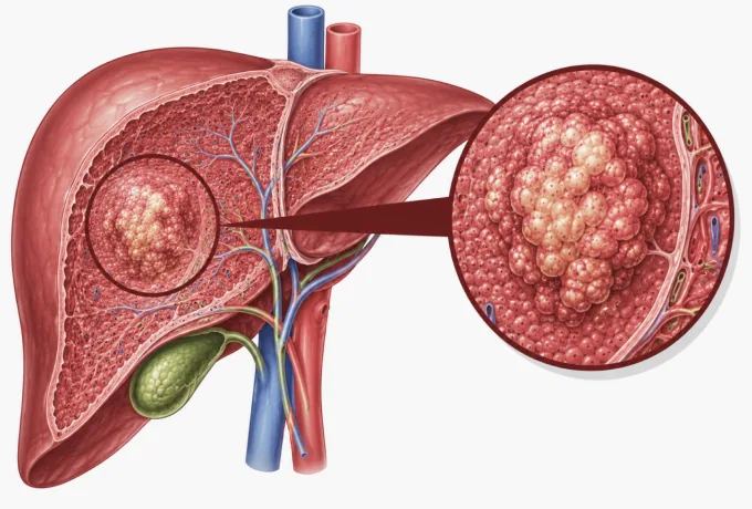
Primary liver cancer begins inside the liver itself — most often in hepatocytes, the organ's main working cells — rather than arriving there from somewhere else. That distinction is not academic: cancer that has spread to the liver from the bowel, breast or lung is a different disease with different treatment, and it is not what this page describes.
What makes liver cancer unusual is the soil it grows in. In most patients the liver was already damaged long before the tumour appeared — by chronic hepatitis B or hepatitis C, by years of heavy drinking, or by metabolic liver disease — and frequently by established cirrhosis. A scarred liver is a liver in which cells are repeatedly injured, lost and replaced, and that cycle of damage and repair is what eventually allows a malignant clone to take hold. The full list is set out in causes and risk factors.
The liver compounds the problem in two ways. Its substance has no pain-sensitive nerves, and it keeps a large functional reserve, so a tumour can grow to a considerable size before producing anything a person would notice. Symptoms are therefore a late signal, not an early one — which is why surveillance imaging, and not symptom-watching, is how this cancer is meant to be found. What to look for if something has already changed is covered in symptoms and early warning signs.
A diagnosis consequently raises three questions that must all be answered before treatment is chosen. How far has the tumour gone? That is the stage. How much working liver is left? That is the functional reserve, usually scored with Child-Pugh. What is driving the tumour? That is the biology — the pathology and molecular findings. Liver cancer care is built on those three axes together, never on the diagnosis alone.
Hepatocellular carcinoma and bile-duct cancer: two diseases, two plans
Most primary liver cancers are hepatocellular carcinoma (HCC), which arises from liver cells. A minority begin instead in the small bile ducts inside the liver and are called intrahepatic cholangiocarcinoma. They are not variations of one illness: they grow differently, are staged differently and respond to different drugs, so the first thing pathology establishes is which one is present.
| Hepatocellular carcinoma (HCC) | Intrahepatic cholangiocarcinoma | |
|---|---|---|
| How common | The large majority of primary liver cancers | A minority of cases, but rising in several regions |
| Where it starts | Liver cells (hepatocytes) | The small bile ducts inside the liver |
| Background liver | Usually already damaged — hepatitis B or C, alcohol, metabolic disease, cirrhosis | Often an otherwise normal liver |
| How it behaves | Often multifocal; tends to invade the portal vein and to stay in the liver longer | Spreads along bile ducts and to lymph nodes earlier |
| Main treatments | Resection or transplant, ablation, TACE and related interventional therapies, radiosurgery, targeted and immunotherapy drugs | Surgery where possible, chemotherapy, targeted therapy, immunotherapy, radiotherapy; interventional treatment in selected cases |
| Targetable findings | Fewer classical driver mutations; immunotherapy combinations are central | More frequent — including FGFR2 fusions and IDH1 mutations in a subset of tumours |
Why the distinction changes the first step. In a patient with cirrhosis, a typical HCC can often be diagnosed from contrast imaging alone, without a needle. Cholangiocarcinoma usually cannot, because its imaging pattern overlaps with other lesions — so tissue is needed. And because a subset of bile-duct cancers carries an alteration with a matching drug, tumour tissue may be tested for those targets. The same patient, with the same sized tumour in the same place, can therefore follow two quite different diagnostic paths depending on which cancer it is.
What the stages mean
Liver cancer is staged differently from most cancers, because the plan has to respect two things at once: the tumour and the liver it sits in. The system most widely used internationally is the Barcelona Clinic Liver Cancer (BCLC) classification, which combines tumour extent, liver function and general fitness. Chinese guidelines use a related national staging system; the underlying logic is the same.
| Stage | What it means | What treatment is usually aiming for |
|---|---|---|
| Very early | A single small tumour in a liver that is still working well | Cure. Removal of the tumour by surgery or ablation, sometimes transplantation |
| Early | One tumour, or up to three, within a size limit; liver function preserved | Cure or long control. Resection, transplantation, or ablation where surgery is not suitable |
| Intermediate | Multiple tumours confined to the liver, without invasion of the portal vein or spread beyond it | Control and conversion. Interventional treatment such as TACE, often repeated, sometimes leading to surgery |
| Advanced | Tumour has entered the portal vein, or has spread to lymph nodes, bone or elsewhere | Long-term control. Targeted therapy and immunotherapy, with radiotherapy or interventional treatment at specific sites when useful |
| End stage | Liver function is failing and the patient's general condition limits treatment | Best supportive care and control of symptoms, with treatment aimed at comfort and quality of life |
Two sentences deserve to be read twice. First, the stage is not decided by the tumour alone: two patients with an identical tumour can receive completely different recommendations because one liver is compensating well and the other is not. Preserving liver function is part of the treatment, not a side consideration. Second, stage is not fixed by the first scan. If a tumour responds to interventional or drug treatment, an initially inoperable cancer can sometimes shrink enough to be removed — which is why a case like this belongs in front of a multidisciplinary panel rather than a single department.
Why liver function and tumour biology matter as much as stage
In most cancers, the stage tells you the most important thing. In liver cancer it tells you only part of it. The first reason is functional: nearly every available treatment takes something out of the liver, whether that is a segment of tissue, a blood supply, or the liver's tolerance for a drug. A plan that destroys the tumour but leaves the patient in liver failure has failed. That is why the same tumour can be treated aggressively in one patient and conservatively in another who has the same stage but poorer reserve.
The second reason is biological. Tumour tissue is examined for features that predict how the disease will behave and which drugs are worth using. In hepatocellular carcinoma, this increasingly means looking for markers of immune responsiveness and for alterations that later-line drugs can target; in bile-duct cancer, tests may look for FGFR2 fusions or IDH1 mutations, which change the treatment entirely. When the first systemic treatment stops working, repeating the question — what is driving this now? — is more useful than switching drugs at random.
Third, blood tumour markers such as AFP and PIVKA-II support the diagnosis and then track response over time, while a liver function panel shows what the liver itself is doing. Read together with imaging, they tell the team whether a treatment is working and whether it is being tolerated. How these tests are put together into a diagnosis is set out in the diagnosis guide, and how they change the treatment plan in the treatment guide.
Why international patients come to China for liver cancer treatment
Four practical reasons come up most often in conversations with patients and their families. None of them is exotic; all of them are consequential.
Volume, and the pattern recognition that comes with it
Close to half of the world's liver cancer cases occur in China, with roughly 900,000 new diagnoses worldwide each year and the largest share of them here. Teams meet this disease daily, in every stage and presentation — including the vascular and multifocal patterns that are seen only occasionally in lower-volume centres.
The whole pathway under one roof
Surveillance ultrasound and AFP testing, multiphase contrast CT and MRI, pathology and molecular testing, an interventional oncology suite, robotic surgery, breath-tracking radiosurgery and systemic therapy all sit inside the same hospital network. Diagnosis and treatment do not have to be assembled across several institutions or countries.
Earlier access to newer drugs
Drug development for liver cancer in China has moved quickly, and combination protocols developed here — of which the best known is camrelizumab with apatinib — are recognised in NCCN, ESMO and CSCO guidelines. Later-line targeted drugs and bispecific antibodies are in routine use, and mutation-matched clinical trials are an option when standard treatment has been exhausted.
One decision, made by seven specialties
Hepatobiliary surgery, interventional radiology, medical oncology, radiation oncology, hepatology, radiology and pathology review the case together and issue a single plan — within 48 hours of your records being complete — rather than a plan that depends on which department you happened to visit first.










