


Leukemia Overview
Leukemia Overview
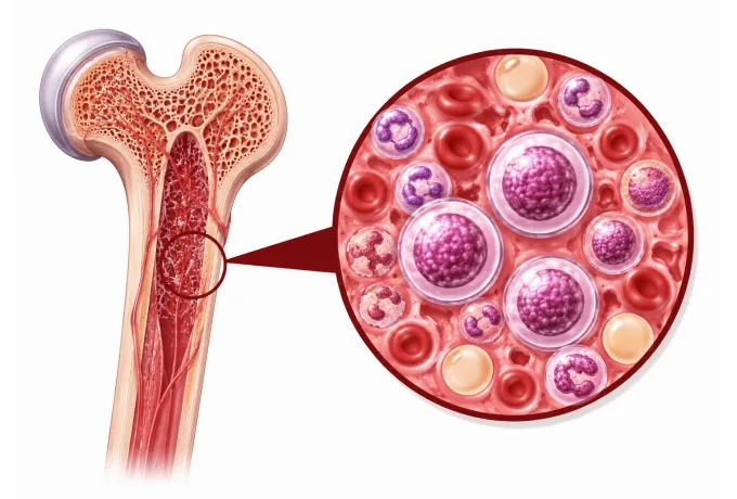
The bone marrow produces every blood cell the body needs: red cells that carry oxygen, platelets that stop bleeding, and white cells that fight infection. Leukemia begins when one of those blood-forming cells acquires the changes that let it multiply without maturing. The abnormal cells do not do the work of the cells they displace — they simply accumulate, and in doing so they crowd out the production of everything else. That mechanism is why leukemia presents as a shortage rather than a lump: too little oxygen-carrying capacity, too few working white cells, and too few platelets.
-
4 types
acute myeloid, acute lymphoblastic, chronic myeloid and chronic lymphocytic leukemia — two axes, not one
-
Marrow
is where it starts: abnormal cells crowd out red cells, white cells and platelets, which is why the symptoms are shortages
-
Film
and flow cytometry are the starting tests; chromosomes and genes confirm the diagnosis and set the risk group
-
48 hrs
from complete records to a personalized MDT plan
What leukemia is
The marrow is a factory that replaces blood cells continuously, and it does so by a sequence of steps: a stem cell divides, its descendants mature, and only then are they released into the blood. Leukemia interrupts that sequence. A cell acquires changes that let it keep dividing without maturing, and because it never leaves the marrow as a working cell, it accumulates there. The marrow becomes filled with cells that are useless rather than with cells that are missing — and the consequences are shortages of everything the marrow should be making.
That gives leukemia its characteristic presentation, which is a combination rather than a single symptom. Too few red cells produce tiredness and breathlessness. Too few working white cells produce infections that are frequent or slow to clear. Too few platelets produce bruising and bleeding — from the gums, the nose, or as pinpoint red spots on the skin. Because the three shortages develop together, patients often describe a set of symptoms that seemed unrelated until a blood count was taken. There is also often no lump at all, which is why leukemia is frequently found by a blood test done for another reason rather than by anything the patient could feel.
The distinction that shapes everything that follows is between acute and chronic. In acute leukemia the marrow is crowded by cells that cannot mature, the disease progresses over days to weeks, and treatment begins as soon as the diagnosis is confirmed — there is no reasonable option to wait. In chronic leukemia the cells retain more of their maturing ability, and the disease behaves very differently: chronic myeloid leukemia is controlled in most patients by an oral targeted drug taken continuously, and early chronic lymphocytic leukemia may need no treatment for years, only monitoring. The same word covers two situations that call for opposite approaches, which is why the type — always stated in full — matters more than the diagnosis on its own.
The four main types
Leukemia is classified along two axes at once, and a full diagnosis names both: how quickly the disease behaves (acute or chronic) and which lineage it arises from (myeloid or lymphoid). The four resulting combinations are the diagnoses most patients are given:
- Acute myeloid leukemia — the commonest acute leukemia in adults, arising from the myeloid line that produces granulocytes, monocytes and platelets. It is treated with combination chemotherapy given as induction followed by consolidation, and the chromosome and gene results decide whether a targeted drug is added and whether a transplant is recommended.
- Acute promyelocytic leukemia — a distinct subtype of acute myeloid leukemia defined by a specific fusion gene, and the exception that proves the rule: it is treated with two non-chemotherapy drugs, all-trans retinoic acid and arsenic trioxide, and it is a medical emergency at presentation because of a clotting disturbance that requires treatment within hours.
- Acute lymphoblastic leukemia — arising from the lymphoid line, the commonest childhood cancer and also a disease of adults, in whom it is less common but harder to treat. The backbone is multi-agent chemotherapy given over two to three years, with a tyrosine kinase inhibitor added when a particular fusion gene is present.
- Chronic myeloid leukemia — defined by the same fusion gene that gives rise to the Philadelphia chromosome, and the disease in which targeted therapy changed the outcome most dramatically. It is controlled in most patients by an oral tyrosine kinase inhibitor, monitored by a blood test that measures the fusion gene.
- Chronic lymphocytic leukemia — an accumulation of mature-looking lymphocytes, most often found incidentally on a blood count in an older adult, and the leukemia that most often requires no immediate treatment at all.
- The rarer types — chronic myelomonocytic leukemia, juvenile myelomonocytic leukemia, hairy cell leukemia, large granular lymphocytic leukemia and blastic plasmacytoid dendritic cell neoplasm, each with its own behaviour and its own small set of effective drugs.
Phases, staging and risk groups
Leukemia is not staged with the tumour-node-metastasis system, because it does not grow as a solid mass. Instead, each type has its own way of describing how far the disease has gone and how urgently it must be treated — and in three of the four types, that description is as important as the diagnosis itself:
| Category | In plain terms | How it is used |
|---|---|---|
| Chronic myeloid leukemia: phase | Chronic phase, accelerated phase, or blast phase — describing how much of the marrow is occupied by immature cells | Determines whether an oral tyrosine kinase inhibitor is expected to control the disease, or whether more intensive treatment and a transplant are needed |
| Chronic lymphocytic leukemia: Rai and Binet stage | From a raised lymphocyte count with no other findings, through enlarged nodes and spleen, to anaemia or a low platelet count | Early stages are monitored rather than treated; the stage is one of several factors deciding when treatment begins |
| Acute myeloid leukemia: risk group | Favourable, intermediate or adverse, assigned from the chromosomes and the gene results | Decides whether chemotherapy alone is likely to be enough, or whether a transplant should be planned in first remission |
| Acute lymphoblastic leukemia: risk group | Based on age, the white cell count at diagnosis, the chromosome findings and the response to the first weeks of treatment | Sets the intensity of the regimen and identifies patients for whom a transplant or cell therapy should be considered early |
| Measurable residual disease | The amount of disease detectable by a sensitive test when the marrow looks normal under the microscope | In acute leukemias it is the single strongest indicator of how deeply treatment has worked, and it can change the plan |
| Response milestones | In chronic myeloid leukemia, the level of the fusion gene measured in the blood at fixed intervals | Defines whether the drug is working, whether the dose should change, and whether a deeper response might allow treatment to stop |
Two features of that table are worth drawing out. In leukemia, the assessment is repeated rather than performed once. Because the disease lives in the marrow and in the blood, its response can be measured directly and repeatedly, and the result at defined time points changes what is given next. This is unlike a solid tumour, where the assessment is an image taken at intervals; here it is a test that can detect disease far below what can be seen under a microscope, which is why measurable residual disease carries so much weight.
The second feature is that the risk group and the treatment are decided together. In acute myeloid leukemia, a favourable-risk result may mean that chemotherapy alone is the plan, while an adverse-risk result usually means a transplant is discussed in first remission — the same diagnosis, and two different treatment strategies, decided by the chromosomes and genes. A plan that states the diagnosis without the risk group is therefore not yet a plan, and asking for the risk group and the reason for it is a reasonable question at the outset.
The markers that guide treatment
In leukemia, testing the chromosomes and genes is not an adjunct to the diagnosis — in several types it is what the diagnosis turns on, and in all of them it determines which drugs can be used. A short list carries most of the decisions:
| Marker | What it is | How it changes management |
|---|---|---|
| BCR::ABL1 | A fusion gene formed by two chromosomes joining — the Philadelphia chromosome — present in almost all chronic myeloid leukemia and in a subset of acute lymphoblastic leukemia | It is the diagnosis in chronic myeloid leukemia and the target of the tyrosine kinase inhibitors that control it; its level in the blood is the measure used to follow the disease |
| PML::RARA | A fusion gene that blocks a specific step in the maturation of myeloid cells | Identifies acute promyelocytic leukemia, which is treated with all-trans retinoic acid and arsenic trioxide rather than conventional chemotherapy — and is a clotting emergency at presentation |
| FLT3 and NPM1 | Two of the most frequent gene changes in acute myeloid leukemia, often occurring together | FLT3 is the target of specific inhibitors added to first-line treatment and used in relapse; NPM1 without adverse features places the disease in a more favourable risk group |
| TP53 and del(17p) | A loss of function of a gene that normally stops damaged cells from dividing | Marks disease that responds poorly to standard chemotherapy, and in chronic lymphocytic leukemia directs treatment away from chemotherapy and towards targeted drugs |
Beyond those four, several other results change what is offered. In acute myeloid leukemia, rearrangements of KMT2A and abnormalities of chromosome 3 or 7 place the disease in a less favourable group, and changes in IDH1 or IDH2 open two further targeted drugs. In acute lymphoblastic leukemia, the presence of BCR::ABL1 or of a Philadelphia-like signature makes a tyrosine kinase inhibitor part of the regimen, while hypodiploidy identifies disease that warrants early consideration of a transplant or cell therapy. In chronic lymphocytic leukemia, IGHV mutation status distinguishes a form that behaves more slowly, and TP53 and del(17p) determine whether chemotherapy has any useful role at all.
The practical instruction is to ask for three things in writing: the full diagnostic name including the subtype, the chromosome and molecular findings, and their effect on the plan. In acute promyelocytic leukemia this is at its most stark — the fusion gene must be confirmed within hours, not days, because treatment starts immediately and differs entirely from any other acute leukemia.
Why patients come to China for leukemia care
Leukemia care depends on four capabilities, and a service either has them or does not: a laboratory that reports molecular results quickly, treatment protocols that are current, cell therapy and transplant infrastructure, and the supportive care that makes intensive treatment survivable.
- Rapid molecular diagnostics — flow cytometry, chromosome analysis and gene testing available in-house and reported within days, because in acute promyelocytic leukemia the result changes treatment to be given that same day.
- Current protocols by subtype — chemotherapy protocols chosen by type and risk group rather than one regimen for all, with targeted drugs matched to the fusion gene or mutation found.
- Cell therapy — CAR T-cell therapy for relapsed or refractory B-cell acute lymphoblastic leukemia, and bispecific antibodies and antibody-drug conjugates that provide an alternative when cell therapy is not suitable.
- Allogeneic stem cell transplant — a transplant programme with unrelated and haploidentical donors, since many patients do not have a matched sibling.
- Supportive care as a discipline — anti-infective prophylaxis, transfusion support, tumour lysis prevention and intensive care, which together determine how much of an intensive regimen a patient can actually complete.
- Measurable residual disease testing — the sensitive tests that show how deeply the disease has responded, and which we use to decide between continuing, changing and proceeding to transplant.
Two practical consequences follow. Because the plan depends on the molecular result, arranging the marrow and molecular testing is the first step — and if the tests were done elsewhere, sending the report together with the slides or the block allows them to be reviewed rather than repeated. And because leukemia treatment is measured in phases with assessments between them, the written plan should state not only the regimen but the time points at which it will be assessed and what each result would change. Send the marrow report, the flow cytometry, the chromosome and molecular results, the treatment given so far and the latest blood counts, and the written assessment is returned within 48 hours of the file being complete.










