


Kidney Stones Overview
Kidney Stones Overview
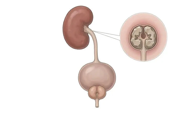
A kidney stone is a crystal that forms when the minerals dissolved in urine — calcium, oxalate, uric acid — become concentrated enough to stop staying dissolved. Many stones cause no trouble at all where they sit in the kidney; the disease announces itself when a stone drops into the ureter, blocks the flow of urine, and produces renal colic — one of the most severe and most characteristic pains in medicine. The treatment side of this condition is genuinely good news: nearly every stone can now be dealt with without a large open operation, through shock-wave lithotripsy, ureteroscopic laser lithotripsy or percutaneous surgery, chosen by the stone's size, position and hardness. The less fashionable half of the plan matters just as much: stones recur, and a stone that is removed without asking why it formed is only half treated. That is why stone analysis and a metabolic evaluation sit at the centre of this pathway, and why the prevention programme — fluid, salt, diet and follow-up — is written down like a prescription rather than offered as advice.
-
A plumbing problem with a chemistry cause
the stone blocks the pipe, but the urine chemistry made the stone
-
Renal colic is the signature
wave-like flank pain radiating to the groin — severe, but rarely dangerous on its own
-
Almost no stone needs open surgery
shock waves, scopes and lasers deal with nearly everything, chosen by size and site
-
48 hrs
from complete records to a personalised MDT plan
Kidney stones: crystals, plumbing and the pain
Urine carries dissolved minerals — calcium, oxalate, uric acid, phosphate — in a solution that is normally kept stable by water and by protective substances such as citrate. When the balance tips, through low fluid intake or an excess of one ingredient, crystals form, grow, and eventually become a stone. Stones sit most often in the collecting system of the kidney: the calyces and the renal pelvis.
While a stone stays in the kidney it is often silent, and a substantial number are found by accident on scans done for other reasons. Trouble begins when a stone drops into the ureter — the narrow tube that drains urine to the bladder. There it obstructs, the kidney swells behind the blockage, and the result is renal colic: pain that arrives in waves, builds to a severity patients describe among the worst they have known, and often radiates from the loin toward the groin.
Two properties of stones shape the whole plan. First, most stones are small enough to pass: with fluid, pain control and sometimes medication, the majority of stones below about half a centimetre leave the body without surgery. Second, the stones that cannot pass can be removed without opening anyone: shock waves from outside the body, a scope through the natural urinary channel with a laser, or a keyhole track into the kidney — matched to the stone rather than chosen by habit.
And one distinction frames the modern pathway: removing the stone answers only half the question. Stones are a metabolic disease that presents as a plumbing emergency. Every stone that passes or is removed is a sample worth analysing, and every patient who has formed one deserves to know why — because the second stone is easier to prevent than to treat.
Stone types: what each stone is made of, and why it matters
Stones are named by their chemistry, and the chemistry is not trivia: it decides both treatment and prevention. The four groups below cover almost everything seen in practice.
| Stone type | Share and chemistry | What it changes |
|---|---|---|
| Calcium oxalate (with calcium phosphate) | By far the commonest group; calcium crystals bound with oxalate | Prevention centres on fluid, salt restriction, normal dietary calcium and oxalate management; often radio-opaque on plain imaging |
| Uric acid stones | Crystals of uric acid, typically in acidic urine; linked with metabolic syndrome and a purine-rich diet | The notable exception: often dissolvable — urine alkalinisation can clear the stone without surgery |
| Struvite (infection) stones | Formed in urine infected with urease-producing bacteria; can grow quickly into a cast of the kidney | Surgical disease: the whole stone must be removed and the infection treated, or it regrows |
| Cystine and rare stones | Genetic disorders of urine chemistry; start early and recur often | Lifelong metabolic management with high fluid targets and specific medication; specialist follow-up |
Position matters alongside chemistry. A stone in a calyx may never move; a stone in the renal pelvis has more room; a stone that has entered the ureter is the one causing colic, and its level — upper, mid or lower ureter — strongly influences both the chance of spontaneous passage and the choice of procedure.
Two axes: the stone itself, and what it is doing to the patient
Stone disease is read on two axes at once. The first describes the stone — its size, its position, how many there are. The second describes the patient — whether the stone is silent, causing colic, obstructing with infection, or affecting kidney function. The second axis decides urgency; the first decides the method.
| Category | Typical picture | What it asks for |
|---|---|---|
| Small stone, no symptoms | Found incidentally or passed already; a stone sitting in a calyx | Imaging follow-up, a fluid and diet programme; intervention only if it grows, blocks or keeps causing trouble |
| Ureteric stone with colic, no infection | Renal colic, blood in the urine, kidney swollen but functioning | Watchful waiting with medical expulsive therapy for small stones; lithotripsy or ureteroscopy for stones unlikely to pass |
| Obstructed kidney with fever | Pain plus fever, chills or feeling profoundly unwell — an infected, obstructed system | An emergency. Drainage first — a ureteric stent or a nephrostomy — then antibiotics, then the stone itself |
| Large or complex stone burden | Stones over about two centimetres, staghorn stones, multiple stones, or stones in abnormal anatomy | Planned percutaneous surgery or a combined strategy, with a metabolic workup built into the pathway |
One honest note belongs here: size alone does not decide the plan. A five-millimetre stone in the lower ureter and a five-millimetre stone in a difficult calyx are different problems, and hardness, anatomy and the patient's own priorities are read together with the measurements.
How the diagnosis is made: imaging, urine and blood
The workup answers four questions in one admission: is there a stone, where is it, what is it doing to the kidney, and — for anyone who has formed stones before — why did it form?
| Step | What it answers |
|---|---|
| Non-contrast CT (CT KUB) | The reference standard: it shows the stone, its exact level, its size and the degree of obstruction, all without contrast |
| Ultrasound | The first-line alternative in pregnancy, in children and for follow-up; it shows swelling of the kidney well and stones imperfectly |
| Urinalysis and culture | Blood in the urine, signs of infection, and the urine pH that clues the chemistry |
| Blood tests | Kidney function, calcium, uric acid and electrolytes; a creatinine that is rising changes the tempo of everything |
| Stone analysis | Whenever a stone is available, from a sieve or from surgery: the composition is the single most useful fact in this disease |
| 24-hour urine studies | For recurrent formers: volume, calcium, oxalate, citrate, uric acid and sodium, measured so the prevention programme targets the actual chemistry |
What the workup should produce is not just a diagnosis but a plan: which stone needs what, when — and what will be done about the next one.
Why patients travel to China for stone treatment
Stone surgery rewards volume and equipment: units that perform ureteroscopy and percutaneous surgery daily operate faster, through smaller instruments, with shorter anaesthetics. The urology departments in our network work within a full endourological armamentarium — flexible and semi-rigid ureteroscopy with holmium laser lithotripsy, shock-wave lithotripsy and percutaneous nephrolithotomy — and within a system built for international patients: records reviewed before travel, imaging and surgical planning completed quickly, and a written plan from a multidisciplinary team within about 48 hours of complete records arriving.
Cost is part of the honesty of the offer: complete treatment here is typically 30%–50% of comparable care in Europe or the US, with more than 30 international insurers working with us on direct billing, and patients from 124 countries and regions. What patients most often tell us matters, though, is the other half of the pathway: that the visit does not end when the stone is gone. Stone analysis and a metabolic evaluation are part of the standard plan, and the prevention programme is written down — with targets for fluid, salt and diet — rather than left as a good intention.










