


Kidney Cancer Overview
Kidney Cancer Overview
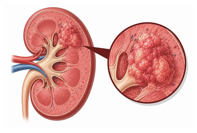
The kidneys sit behind the abdominal organs, filtering blood continuously and producing urine without any sensation the patient can detect. A tumour growing inside one of them therefore has nothing to press against and nothing to bleed from that would announce it. That anatomy explains the single most important fact about this cancer: more than half of kidney tumours are now found by accident, on a scan performed for an unrelated reason.
-
About 90%
of kidney cancers are renal cell carcinomas, arising from the filtering tubules
-
About 75%
of those are the clear cell subtype, which behaves differently from the others
-
More than half
are found incidentally on imaging done for another reason
-
48 hrs
written multidisciplinary plan after the file is complete
What kidney cancer is
Each kidney contains roughly a million nephrons — microscopic tubes that filter blood and produce urine. The great majority of kidney cancers begin in the cells lining those tubes, which is why they are called renal cell carcinomas and why their behaviour differs so much from cancers that start in the drainage system rather than the filtering tissue.
That distinction matters for a reason patients meet immediately. A tumour in the filtering tissue grows outward into the kidney substance and can be removed while leaving the rest of the kidney intact. A tumour in the collecting system — the renal pelvis — is a different disease, called upper tract urothelial carcinoma, and is treated differently. Both can cause blood in the urine; only imaging reliably separates them, and the treatment discussion should make clear which one is being described.
The anatomy also explains why symptoms are a poor guide. The kidneys are wrapped in fat and lie against the back muscles, with no organ close enough to be compressed by a small tumour. There is no bowel to obstruct, no duct to block at an early size, and no surface to bleed readily. By the time a kidney tumour produces pain or a palpable mass it has usually grown well beyond the organ — which is why imaging, not symptoms, is what finds this cancer early, and why more than half of tumours are now discovered while looking for something else.
One structural feature shapes treatment throughout: a person has two kidneys, and losing one is survivable but not free. The remaining kidney has to take over the whole filtering load, and if its function was already reduced, removing the other organ can push someone toward dialysis. That is the reasoning behind the most important trend in this cancer over the past two decades — preserving as much kidney as the tumour permits, rather than removing the whole organ by default.
The main subtypes
Kidney cancer is not one disease, and the subtype is established by the pathologist from the tissue, not guessed from the scan. The distinction matters because the subtypes respond differently to the drugs used in advanced disease:
- Clear cell renal cell carcinoma — about three quarters of cases. Named for the pale cytoplasm seen under the microscope, which reflects stored lipid and glycogen. It is the subtype around which most immunotherapy and targeted treatment evidence is built.
- Papillary renal cell carcinoma — around one in seven. It forms finger-like projections and is divided into type 1 and type 2, which behave differently. A subset is driven by changes in the MET gene, which opens a specific targeted option.
- Chromophobe renal cell carcinoma — around one in twenty. It tends to grow slowly and is graded by different criteria from the other subtypes, which is why a grade given for one subtype should not be read across to another.
- Rare subtypes — collecting duct carcinoma, renal medullary carcinoma, translocation-associated carcinoma and unclassified tumours. These are uncommon but often behave more aggressively, and several are linked to inherited conditions, so their presence should prompt a genetics discussion.
Two practical consequences follow. First, treatment for advanced disease now depends on the subtype as much as on the stage, so a pathology report should state the subtype explicitly rather than only the word carcinoma. Second, certain subtypes — particularly those occurring in young patients, in both kidneys, or with a family history — are signals of an inherited syndrome, which changes surveillance for the patient and testing for relatives. Both are questions worth putting to the treating team in writing.
What each stage means
Staging uses the AJCC system, which describes where the tumour is: inside the kidney, into the fat around it, into the vein that drains it, or beyond. Treatment of localised disease turns on the size and position of the tumour; treatment of advanced disease turns on the risk group rather than the stage number:
| Category | In plain terms | How it is usually approached |
|---|---|---|
| T1a — up to 4 cm | A small tumour confined to the kidney | Partial nephrectomy where feasible; ablation or active surveillance are options in selected patients |
| T1b — over 4 to 7 cm | Confined to the kidney, but larger | Partial nephrectomy where technically feasible; otherwise radical nephrectomy |
| T2 — over 7 cm | Confined to the kidney, but large | Radical nephrectomy, with kidney-preserving surgery considered case by case |
| T3 — locally advanced | Into the fat around the kidney, the renal vein, or the large vein above it | Surgery, sometimes with a venous thrombus removed at the same time; systemic treatment in selected cases |
| T4 — beyond the kidney | Into the adrenal gland or beyond the outer fascia | Surgery where it can be complete, combined with systemic treatment |
| N and M | N: cancer in lymph nodes. M: spread to lung, bone, liver or elsewhere | Systemic treatment chosen by risk group and subtype, with surgery reserved for selected situations |
Two features of that table repay attention. Size is doing more work than stage letters in early disease: the line between a 4 cm and a 5 cm tumour is the line between a straightforward partial nephrectomy and a more difficult decision, which is why the measurement is worth confirming before the operation is planned. And a venous thrombus is not the same as spread: a tumour growing into the renal vein can still be removed completely, which is why the imaging report must state how far up the vein it extends.
Markers that guide treatment
For advanced kidney cancer the choice of drug is no longer made by stage alone. Four pieces of information drive it:
| Marker or group | What it tells the team |
|---|---|
| Histological subtype | Clear cell tumours follow the main immunotherapy and targeted evidence; papillary and chromophobe tumours are treated differently, and MET-driven papillary tumours have a specific targeted option |
| IMDC risk group | Six routine factors — performance status, time from diagnosis to treatment, haemoglobin, calcium, neutrophils and platelets — place the disease in a favourable, intermediate or poor group, which is what decides whether an immunotherapy combination or a single targeted drug is used first |
| PD-L1 and related markers | Support the decision between an immunotherapy combination and a targeted single agent, but are not on their own decisive in kidney cancer |
| Germline testing | Indicated for young patients, bilateral or multifocal tumours, a family history, or certain subtypes — and can change surveillance and screening for relatives |
The practical point is that these are not optional extras layered on top of a treatment decision; they are the decision. A pathology report that states only the cancer type leaves the advanced-disease conversation without its starting point, and the risk group cannot be calculated without the blood results and a performance assessment. This is also where a written records review adds most value, because it can say which single missing item would change the recommendation.
Why patients come to China
Kidney cancer care turns on four capabilities, and a centre either has them or does not: an accurate scan read the same way twice, a surgeon who will preserve the kidney whenever the tumour allows, ablation and surveillance offered as genuine alternatives rather than afterthoughts, and systemic treatment that keeps pace with a field moving quickly.
- Kidney-preserving surgery — partial nephrectomy, including robot-assisted and laparoscopic approaches, planned from the scan so that the amount of kidney to be spared is decided before the operation rather than during it.
- Complex and locally advanced surgery — tumours extending into the renal vein or the large vein above it, which require a planned vascular approach rather than an improvised one.
- Ablation and surveillance as real options — thermal ablation for small tumours in patients who cannot have surgery, and structured active surveillance with a defined imaging interval for those whose tumour is unlikely to cause harm within their lifetime.
- Subtype-aware systemic treatment — immunotherapy combinations, targeted agents and HIF-2 inhibition for advanced disease, chosen from the subtype and risk group rather than from stage alone.
- Kidney function watched throughout — creatinine and estimated filtration rate tracked before and after treatment, because preserving function is part of the goal and not a side consideration.
- Genetics where it changes management — referral and testing for patients whose age, family history or subtype suggests an inherited syndrome.
Two practical conclusions follow. Because the choice between surveillance, ablation and surgery depends on tumour size and position, on kidney function and on the patient’s own priorities, a written assessment should set out all three routes rather than present one as settled. And because kidney function is a shared concern across every route, the creatinine and filtration results belong in the first set of records sent. Send the multiphasic CT images, the pathology report and the recent blood tests together, and a written assessment follows within 48 hours of the file being complete.










