


Ischaemic Stroke Overview
Ischaemic Stroke Overview
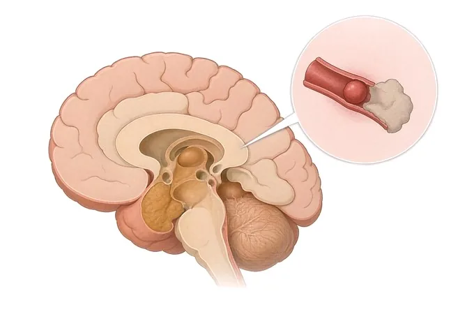
An ischaemic stroke is what happens when an artery supplying part of the brain is blocked and the tissue downstream begins to die. It is the commonest form of stroke, it is a medical emergency rather than a neurological curiosity, and almost everything about the outcome depends on how quickly the blockage is found and how much of the brain can still be saved. This guide follows one thread throughout: the same blocked artery asks two separate questions — whether it can be opened, and what must be done afterwards so that it does not happen again.
-
24/7
Stroke thrombolysis and thrombectomy cover
-
48 hours
Written second-opinion report after records arrive
-
30+
International insurers working with us on direct billing
-
124+
Countries and regions our international patients come from
What an ischaemic stroke is — a blocked artery, not a burst one
A stroke is a sudden loss of brain function caused by a problem with the brain's blood supply. In the ischaemic form — roughly four strokes in five — an artery is blocked, and the brain tissue it supplies stops receiving oxygen and glucose. Neurones are the most oxygen-dependent cells in the body and the least able to store energy, so they begin to fail within minutes. The centre of the affected territory, the core, is lost quickly. Around it lies a rim of tissue that is not receiving enough blood to function but is still alive, kept going by other vessels: the penumbra. The whole purpose of acute stroke treatment is to reopen the artery before the penumbra becomes core.
This is why an ischaemic stroke is not a single event that is simply over when the symptoms stop worsening. It is an evolving process that continues for hours, and every minute without flow costs brain. The figure most often quoted in stroke medicine is that around two million neurones are lost for every minute that a large artery stays blocked. Whether that number is precisely right matters less than what it implies for practice: the question "when was the patient last known to be well?" is asked before almost any other, because it is the answer that decides what can be offered.
The boundary between ischaemic and haemorrhagic stroke is not academic. In a haemorrhagic stroke a vessel has burst and blood is escaping into or around the brain. Both produce sudden weakness, speech difficulty or loss of vision, and neither can be told from the other by examination. The first test is therefore a non-contrast CT of the head, not a sophisticated one — because clot-dissolving treatment and clot retrieval are appropriate for a blocked artery and actively harmful for one that has bled. The distinction is also the reason this guide is separate from the one on cerebral haemorrhage: the two conditions share their symptoms and almost none of their treatment.
Why the mechanism matters more than the label
"Stroke" describes a consequence, not a cause, and two patients with identical weakness on the same side may need entirely different treatment for years afterwards. The classification used in practice groups strokes by how the artery came to be blocked, because that determines what can be changed and what must be watched. The five groups below cover almost every patient, and establishing which one applies is as much a part of the work-up as the scan itself.
| Mechanism | What is blocking the artery | What it means for treatment |
|---|---|---|
| Large-artery atherosclerosis | An atherosclerotic plaque in a carotid or intracranial artery has narrowed it, and a clot has formed or travelled onto the plaque | Long-term antiplatelet treatment and intensive risk-factor control; a symptomatic narrowing in the neck may call for endarterectomy or stenting. The emergency treatment depends on whether a large vessel is occluded |
| Cardioembolism | A clot has formed in the heart and travelled to the brain — most often in atrial fibrillation, also in valve disease, after a heart attack, or with a hole in the heart | Anticoagulation rather than antiplatelet treatment, started once it is safe in relation to the size of the infarct. Finding an irregular rhythm that has never been documented changes the plan for decades |
| Small-vessel occlusion | Disease of the small penetrating arteries deep in the brain, usually on a background of high blood pressure, diabetes and smoking | No large artery to open. Treatment is risk-factor control — blood pressure above all — plus antiplatelet medication and lifestyle change |
| Other determined cause | Arterial dissection, vasculitis, a clotting disorder, a blood disorder, drug use, or a complication of pregnancy and the postpartum period | Treatment follows the cause and is often different from routine stroke care: anticoagulation for dissection, immunosuppression for vasculitis, condition-specific management in pregnancy |
| Undetermined cause | Thorough investigation finds no definite mechanism — the group called cryptogenic, and a substantial share of strokes in younger adults | Treatment is decided from what remains plausible. Prolonged rhythm monitoring and a search for a hole in the heart are the two investigations most often added later |
A second classification matters for the emergency phase, and it is anatomical. A stroke in the carotid territory — the middle cerebral artery and its branches — typically produces weakness and sensory loss on one side, a turned face, and difficulty with language. A stroke in the vertebrobasilar territory, supplying the brainstem, cerebellum and the back of the brain, produces dizziness with unsteadiness, double vision, difficulty swallowing or speaking clearly, and loss of one side of the visual field. The second is the more often missed, because none of its features is weakness, and because the word "dizziness" is used for a great many conditions that are not stroke.
How a stroke is measured: NIHSS, the occlusion site and the salvageable brain
Two questions are asked together and must be kept apart. The first is how much brain is already lost, and how much is still at risk — answered by imaging. The second is how disabled the patient is right now — answered by a clinical score. The first decides whether reopening the artery is still worth attempting; the second describes the patient, and it can move in either direction within hours.
| Measure | What it captures | Why it changes the decision |
|---|---|---|
| NIHSS score | A bedside scale from 0 to 42 covering consciousness, gaze, visual fields, movement, sensation, speech and coordination | It grades severity and it is repeated — a rising score is an emergency. It is not a treatment threshold on its own, and it under-weights posterior-circulation strokes |
| Occlusion site | Where the artery is blocked: internal carotid, main trunk of the middle cerebral artery, a branch, or the basilar artery | This is the single most important predictor of what can be achieved by removing the clot; a proximal large-vessel occlusion is the target of thrombectomy, and a distal branch occlusion usually is not |
| ASPECTS | A ten-point score read from the CT, subtracting a point for each region of the middle cerebral territory already showing early ischaemic change | A low score means much of the territory is already lost, which makes reopening the artery less likely to help and raises the risk of bleeding into the infarct |
| Core and penumbra | The volume of tissue that is already dead, compared with the volume that is hypoperfused but still salvageable, measured by CT or MRI perfusion | This is what allows treatment beyond the standard window: a small core with a large penumbra is the pattern in which late thrombectomy still helps |
| Time from last known well | Time from the moment the patient was last known to be without symptoms — not from the moment they were found | It sets which treatments are available. It is also the reason waking with symptoms is treated as an unknown-time stroke until imaging shows otherwise |
| Modified Rankin Scale | A six-point description of functional independence, from no symptoms to death | The primary outcome by which treatment is judged, which is why it is recorded at presentation rather than on discharge only |
Three consequences of this table are worth stating plainly. A low NIHSS does not mean a mild problem: a small infarct in the brainstem can leave a patient unable to swallow, walk or speak clearly, while a larger infarct in a silent region can produce very little. A high NIHSS does not mean treatment is futile: it is often the sign of a large-vessel occlusion, which is precisely the situation thrombectomy was developed for. And the score and the scan answer different questions: the score describes the patient, the scan describes the brain, and treatment decisions require both.
What each test answers in the first hours
The tests are done in a deliberate order, and the order is designed so that nothing delays the decision about treatment. Each of them answers a different question, and none of them is a substitute for another.
| Test | The question it answers | What it cannot do |
|---|---|---|
| Non-contrast CT of the head | Is this bleeding or is it a blocked artery? It is fast, universally available and it excludes haemorrhage in the great majority of patients | It is normal or near-normal in the first hours of an ischaemic stroke, so a normal scan does not exclude the diagnosis |
| CT angiography | Where exactly is the blockage, is a large vessel occluded, and what do the arteries of the neck look like? | It shows anatomy rather than viability — an occluded artery does not by itself tell you whether the tissue behind it can still be saved |
| CT or MRI perfusion | How much brain is dead already, and how much is at risk? This is what identifies patients who can still benefit after the standard time window | It requires software and experience to interpret; thresholds differ between centres and should not be applied by numbers alone |
| MRI including diffusion imaging | The most sensitive way to demonstrate infarction, and the way small and posterior-circulation strokes are found | It is slower and less available in an emergency, and it cannot be done in some patients; it does not replace the first CT |
| Cardiac investigation | Is there atrial fibrillation or another cardiac source of clot — electrocardiogram, prolonged rhythm monitoring and echocardiography | A normal electrocardiogram does not exclude a rhythm problem: paroxysmal atrial fibrillation is intermittent by nature and needs monitoring to be caught |
| Blood tests and vascular imaging | Whether there is a treatable cause and what the risk profile is: glucose, lipids, clotting, and imaging of the carotid and intracranial arteries | They do not change emergency treatment; with rare exceptions they belong to the hours after reperfusion has been decided |
Why patients travel to China for stroke care
Acute stroke treatment is standardised worldwide, and no one should travel during the first hours of a stroke — the treatment that matters most is the one given in the nearest hospital that can provide it. Where travel does make sense is in the two situations that follow: establishing the cause when the mechanism remains unclear, and rehabilitation, which is where most of the recovery is made and where time and continuity matter more than equipment.
Our neurology and neurosurgery services are organised as a standardised stroke centre built to national stroke-centre standards, with intravenous thrombolysis and endovascular thrombectomy provided as bridging therapy and whole-process management, and thrombolysis and thrombectomy cover available around the clock. Cerebrovascular cases are planned jointly by neurology, neurosurgery and rehabilitation rather than handed from one to the next, and interventional work — including mechanical thrombectomy, stent implantation and the treatment of aneurysms, arteriovenous malformations and arterial stenosis — is carried out on 3D DSA, alongside 3.0T MRI, dual-source CT, PET-CT and 24-hour video EEG. Neurological rehabilitation is integrated into the same pathway as the acute treatment rather than offered as a separate service afterwards.
The practical questions are answered in the same way as for any other condition we treat. Records reviewed by a stroke specialist receive a written opinion within about 48 hours of complete records arriving, and the opinion states the mechanism if it can be established from the material, what remains uncertain, and which investigations would settle it. Complete treatment in China typically runs at 30–50% of comparable care in Europe or the United States. We work with more than 30 commercial insurers for direct billing and have received patients from 124 countries and regions. Where the question is one that can be answered from images and records alone, a video consultation is often the sensible first step rather than a journey made to ask it.










