


IgA Nephropathy Overview
IgA Nephropathy Overview
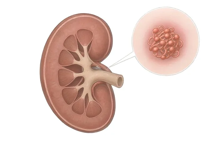
IgA nephropathy is the most common primary glomerular disease found on kidney biopsy worldwide, and it is diagnosed in only one way: by taking a piece of the kidney and looking at it. An abnormal form of immunoglobulin A deposits in the filtering units, the immune system reacts, and the outward sign is blood in the urine — classically within a day or two of a sore throat. This guide follows the path patients actually walk: what the blood and the protein in the urine each mean, how a biopsy is read and scored, what is known about why it happens, and which treatments change where the disease ends up.
-
Biopsy, not blood
the only test that makes the diagnosis
-
1–2 days
the gap between a sore throat and visible blood — not the two to three weeks of a post-streptococcal nephritis
-
Protein, not blood
what sets the long-term risk
-
2004
the year our nephrology department was established
What IgA nephropathy is — an antibody that ends up in the filter
Immunoglobulin A is the antibody the body stations at its moist surfaces — the lining of the nose, mouth, throat and gut — where it is the first thing a virus or bacterium meets. In IgA nephropathy, a structurally abnormal form of IgA1 is produced in larger amounts than usual. Its sugar side-chains are incomplete, the body eventually recognises it as foreign and makes antibodies against it, and the resulting immune complexes circulate until they lodge in the mesangium — the scaffolding at the centre of the kidney's filtering units.
Once deposited, they provoke inflammation. The filter becomes leaky, and what leaks is first red blood cells and later protein. That is the whole of the disease in one sentence: an antibody made at a mucosal surface ends up in the kidney filter, and the filter leaks. Everything a patient notices follows from that, and so does everything we measure.
Two consequences of this mechanism are worth stating at the beginning, because they shape every conversation that follows. First, the diagnosis is histological. The abnormality is where the antibody is, not how much of it is in the blood — the serum IgA level is normal in most people with the disease, and measuring it is not part of the diagnosis. Second, the disease is not one event but a tendency. Each mucosal infection can provoke another round of complex formation, which is why episodes of visible blood keep coming back over years, and why the long-term picture is measured in decades rather than weeks.
The two things that appear on a urine test mean quite different things, and conflating them is the commonest source of both false reassurance and unnecessary alarm. Haematuria — blood — is what a patient sees, and an episode of it does not by itself damage the kidney. Proteinuria — protein — is invisible, and it is the finding that tracks with progressive scarring. A person whose urine clears of blood but whose protein climbs is doing worse, not better. That distinction is why the symptoms page spends more space on what protein means than on what blood looks like.
Primary and secondary forms, and why the difference changes the treatment
IgA deposits are not always the whole story. Sometimes they are the disease; sometimes they are the kidney's response to something else that is going on, and in that case the treatment belongs to the other thing. Separating the two is not an academic exercise — it decides whether the useful medicine is a blood pressure drug, a gluten-free diet, or an antiviral.
| Form | What is driving it | How it usually presents | What it means for treatment |
|---|---|---|---|
| Primary IgA nephropathy | No other disease found; the abnormal IgA1 production is the problem itself | The classic picture — visible haematuria timed with mucosal infections, in a young adult, often found after years of silent microscopic haematuria | The full pathway: optimised supportive care first, disease-targeted and immune therapy only where the risk justifies it |
| Associated with chronic liver disease | The liver normally clears IgA complexes; cirrhosis leaves them in the circulation | A person with known liver disease and a rising creatinine or new proteinuria; often silent haematuria rather than dramatic visible blood | Treat and monitor the liver disease; the kidney management is supportive, and immune suppression is rarely the answer |
| Associated with coeliac disease | Gluten-driven mucosal immune activation with IgA complex formation | Subtle: often found because a clinician looked, sometimes with gastrointestinal symptoms that were attributed elsewhere | A gluten-free diet is the treatment; tested for, not assumed, but worth testing in everyone |
| Associated with inflammatory bowel disease | Chronic intestinal inflammation driving the same mucosal antibody response | Haematuria and proteinuria in a person with established Crohn's disease or ulcerative colitis | Control the bowel disease; the kidney usually follows it |
| Associated with chronic infection | Persistent antigenic stimulation — viral hepatitis, HIV, and some chronic bacterial infections | A person with a known chronic infection in whom the urine changes | Treat the infection; immune suppression without doing so makes matters worse |
| IgA vasculitis with nephritis | The same antibody depositing systemically — in skin, gut and joints as well as kidney | A purpuric rash on the legs and buttocks, abdominal pain, joint pain, with haematuria alongside | A systemic disease managed as one; kidney involvement graded by the same biopsy logic, and usually more aggressive up front |
Notice how few rows end in "give immunosuppression". The screening for a secondary form — liver function, coeliac serology, a careful history of bowel symptoms, hepatitis and HIV serology — is cheap, non-invasive and frequently changes the plan, which is why the diagnosis page puts it before the discussion of immune therapy rather than after.
How risk is graded: the Oxford score and the two numbers that are followed
IgA nephropathy has no stage number that a patient carries around. Instead, severity is graded once from the biopsy and then tracked with two measurements that are repeated for the rest of the patient's life. Understanding both halves makes the follow-up appointments legible instead of mysterious.
The biopsy is scored with the Oxford classification — MEST-C — which reads five features under the microscope. It is not a grade from one to four; it is a profile, and each letter answers a separate question.
| Score | What the pathologist is looking at | What it tells us |
|---|---|---|
| M — mesangial hypercellularity | How many cells are packed into the mesangium, scored M0 or M1 | The burden of deposited complexes. It confirms the disease rather than deciding its fate, and it is the least prognostic of the five |
| E — endocapillary proliferation | Whether inflammatory cells have moved into the capillary loops, E0 or E1 | Active inflammation that has not yet scarred — the feature most likely to respond to anti-inflammatory treatment |
| S — segmental glomerulosclerosis | Scarring confined to part of some filtering units, S0 or S1 | Damage that will not reverse. Its presence argues for protecting what remains rather than expecting recovery |
| T — tubular atrophy and interstitial fibrosis | Scarring of the tubules and the tissue between them, T0, T1 or T2 by extent | The strongest single predictor of the long-term outcome. It is the measure of how much kidney has already been lost, and it cannot be recovered |
| C — crescents | Whether cells have proliferated into the urinary space — C0, C1 or C2 by the proportion of glomeruli involved | Aggressiveness. Many crescents means a rapidly progressive course that needs an urgent and different response |
The score is read once. What happens afterwards is simpler and it is what follow-up is actually for: protein in the urine and filtration rate in the blood, measured at intervals, with blood pressure alongside. The trajectory of those two numbers over one to two years tells us more than the biopsy ever will about where a particular patient is heading, and it is also what tells us whether treatment is working.
| Where the risk sits | What the numbers look like | What we do about it |
|---|---|---|
| Low | Minimal protein, normal filtration, normal blood pressure | Observation with periodic measurement. No immune therapy; the risk of treatment exceeds the risk of the disease |
| Moderate | Persistent protein despite supportive care, filtration still well preserved | Supportive care pushed to its full extent first — this is where most of the lifetime benefit comes from |
| High | Substantial protein that does not settle, falling filtration, or a biopsy with extensive scarring or crescents | Disease-targeted therapy and immune treatment enter the discussion, weighed against infection risk and against how much function is left to protect |
| Established advanced disease | Filtration already severely reduced, with scarring throughout | Immune therapy generally stops being useful. Care shifts to managing the complications of reduced function and planning replacement therapy in good time |
Immunosuppression is not a treatment for advanced disease; it is a treatment for inflammation that is still costing function. Where scarring dominates, a different plan applies — set out on the treatment page.
What a full assessment contains
The assessment exists to answer four questions in order: is this the kidney's filter bleeding rather than the plumbing, is it IgA, is there a secondary cause, and how much kidney has already been lost. Everything on this list serves one of those four.
| Test | What it measures | What it answers |
|---|---|---|
| Urine microscopy | Whether the red cells are dysmorphic, and whether there are red cell casts | The first and cheapest separator: misshapen cells and casts mean the blood came through the filter, which is a nephritis, and not from a stone or a tumour |
| Protein quantification | Protein-to-creatinine ratio on a spot sample, or a 24-hour collection | The number that carries the prognosis, and later the number that tells us whether treatment is working |
| Serum creatinine and filtration rate | How much filtering capacity remains, estimated from the blood | Where on the trajectory the patient is now. A single reading is a snapshot; three readings over six months are a trend, and the trend is what matters |
| Kidney biopsy with immunofluorescence | Light microscopy for the scarring pattern, immunofluorescence for dominant mesangial IgA, electron microscopy for deposit location | The diagnosis itself, plus the Oxford score. This is the one test that cannot be replaced |
| Secondary-cause screen | Liver function and serology, coeliac serology, hepatitis and HIV serology,autoantibodies and complement | Whether the IgA deposition is the disease or a consequence of another one — a distinction that changes the treatment more than anything else on this list |
| Blood pressure, measured properly | Office readings plus home or ambulatory measurement | Both a cause of progression and the most treatable one. Home readings are more informative than a single surgery measurement |
Two practical points. The biopsy is the one test that cannot be replaced: without it, treatments that carry real risk are being given on an assumption. And the protein measurement should be repeated rather than taken once — a single value during an infection or after heavy exercise is misleading.
Why patients come to China for IgA nephropathy
Our nephrology department was established in 2004 and is the leading unit of the Fosun Health Nephrology Specialty Alliance, led by Associate Professor Liu Xiaogang. Primary and secondary kidney disease — IgA nephropathy, diabetic nephropathy, SLE and lupus nephritis among them — is the department's core work rather than a subspecialty of it, and the pathology, the wards and the dialysis unit sit inside the same service.
The department runs an outpatient clinic, inpatient wards and a haemodialysis unit with 20 specialised beds, more than 30 haemodialysis machines and 2 CRRT machines, and handles nearly 20,000 kidney outpatient visits and close to 1,000 inpatient admissions a year. Its equipment — Fresenius haemodialysis machines, haemodiafiltration machines, CRRT, a secondary reverse osmosis water treatment system, a haemodialysis centre information management system and a centralised fluid supply system — is what supports the patients whose disease has already reached the point of replacement therapy.
For the rare aggressive presentations there is also a capability that is not available everywhere: double plasma exchange and hybrid blood purification, alongside peritoneal dialysis catheter placement and vascular access establishment and maintenance. In a rapidly progressive IgA nephropathy this is the difference between having a rescue option in the same building and waiting for a transfer.
There is also an integrated pathway. The traditional Chinese medicine department runs a dedicated kidney disease clinic, and patients who want a combined approach are managed across both services: blood pressure, proteinuria and filtration managed conventionally, with an individualised Chinese medicine regimen alongside it. Where a patient asks about this, the honest framing is that it is an addition rather than an alternative — the protein number that has to come down is still measured in the same units, and the medicines that lower it are still the same medicines.
What international patients mostly come for is the thing a short outpatient appointment rarely produces: a biopsy read and scored, a secondary cause actively excluded, and supportive care maximised before anyone reaches for immunosuppression. The first of those — reviewing the records you already have — happens before any travel is discussed.










