


Hydrocephalus Overview
Hydrocephalus Overview
Hydrocephalus is not water on the brain. It is a problem of flow : the fluid that bathes the brain and the spinal cord is produced, moves and is absorbed in a continuous circuit, and where that circuit is blocked, or where absorption fails, the ventricles enlarge and pressure is transmitted to the tissue around them. That is what makes this condition unusual in neurology — it is mechanical, and in most of its forms it has a mechanical answer. It has three faces , and separating them is most of the work: obstructive, in which the pathway is blocked; communicating, in which absorption has failed; and normal pressure hydrocephalus in adults, which presents as a triad rather than as pressure. A fourth appearance has to be named and excluded rather than treated: ventricles that are large because the brain around them has shrunk are not hydrocephalus at all. This guide follows the order people need it in: what the condition is, the shapes and what separates them, what changes the timescale, how the assessment is assembled, and what treatment actually consists of — which includes the second half that families are told about least often.
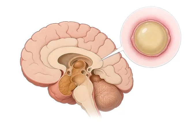
-
48 hours
written opinion once complete records arrive
-
One team
neurology, neurosurgery, imaging, rehabilitation and neuropsychology together
-
A test, not a scan
removing fluid and measuring gait is what predicts benefit
-
124+
countries and regions our international patients come from
What hydrocephalus is
The fluid that surrounds the brain and the spinal cord is not standing water. It is produced continuously, it moves along a set pathway through a system of connected spaces, and it is absorbed continuously back into the circulation. Hydrocephalus is what happens when that circuit goes wrong: either the pathway is obstructed, or absorption fails, and the consequence is that the ventricles — the spaces at the centre — enlarge, and the tissue around them is pushed on from the inside.
That mechanism is worth having clearly, because it explains two things that otherwise look contradictory. It explains why this condition can present with the features of raised pressure in one person and with none of them in another — the same enlargement can occur at a pressure reading that is entirely normal. And it explains why this is one of a small number of neurological conditions in which treatment can genuinely reverse what is happening, rather than only managing it: a mechanical problem has a mechanical answer.
The name itself does the condition a disservice. Water on the brain suggests something pooled and static, and a great many families arrive having understood it that way. What is actually happening is a circulation failing at one point, and which point it fails at is what the assessment exists to establish. It is also worth saying what hydrocephalus is not: it is not a disease of the brain substance, it is not a dementia, and — in the commonest adult form — it is not accompanied by the headaches people expect from pressure.
What matters most from here is not how large the ventricles are. It is which shape this is, because the shapes differ in what treatment can offer and in whether a device is ever the right question. What the condition looks like in a day, and in an infant, is set out on the symptoms page.
The shapes, and what separates them
The table below sets out the shapes that account for most of what presents, and for each one what identifies it and what the distinction changes. The last row is the one to read twice, because it is the one that is treated when it should not be.
| Shape | What has gone wrong | Who and how it presents | What the distinction changes |
|---|---|---|---|
| Obstructive hydrocephalus | The pathway itself is blocked at a point — a narrowing, a cyst, or a mass at any point in the circuit | Pressure features: headache worst in the morning, vomiting, blurred vision, and in an infant a head growing across centiles | The cause is the question rather than the consequence. Where the block can be relieved, the device may not be needed |
| Communicating hydrocephalus | The pathway is open, but absorption at the far end has failed — most often after bleeding, infection or injury | Weeks to months after the event that caused it, with the same pressure features, or with a slowing and a change in a person already unwell | A device is usually the answer. What changes is the timing, and whether the underlying event has left anything else to treat |
| Normal pressure hydrocephalus | Not fully understood: absorption is impaired, but pressure readings are within the usual range | An older adult with gait first, the bladder next, and thinking slowing last. Headache is often absent | It is the most treatable of the three and the most often missed. The gait is the element most likely to respond |
| Congenital and infantile hydrocephalus | Present before birth, most often from a structural cause in the pathway or as a consequence of prematurity | A head growing across centile lines, a full fontanelle, irritability, poor feeding, and eyes resting with the white visible above the iris | Time is tissue rather than time is brain. The measurement that matters is the head plotted over time, not judged by eye |
| Post-haemorrhagic and post-infective hydrocephalus | Absorption damaged by blood or by infection in the spaces around the brain | During recovery from a bleed, a meningitis or a head injury, with a slowing or a failure to progress as expected | Expected rather than surprising after those events. The error is attributing the slowing to the original illness |
| Ventricles large from loss of volume | The tissue around the ventricles has shrunk, and the spaces have enlarged passively to fill it | An older adult with a progressive cognitive decline and no gait of the hydrocephalic shape, no bladder urgency, and no pressure features | Not hydrocephalus, and shunting it does nothing. It adds a device and its risks without changing the course |
Two rows carry most of the consequence in practice. Normal pressure hydrocephalus is the one most often missed, because the triad is read as three separate problems belonging to old age rather than as one condition — and it is the one in which treatment is most likely to give something back. Ventricles enlarged by loss of volume is the one most often over-treated, because the images look alike and the distinction is made on proportion rather than on size. How that distinction is made is set out on the diagnosis page.
What changes the timescale
Two different clocks run in this condition, and confusing them is the commonest way something urgent is allowed to wait. The first is the slow clock of normal pressure hydrocephalus, which is measured in months and in which nothing needs to happen tonight. The second is the clock of a blocked circuit or a failing device, which is measured in hours. What follows separates them, with the action attached to each.
| Feature | What it looks like | What it warrants |
|---|---|---|
| Headache with vomiting, or drowsiness | Morning headache, vomiting without nausea, increasing sleepiness, or a person who is difficult to rouse | Assessment now, not at the next appointment. This is the pattern of pressure rising rather than of a condition progressing |
| A symptom returning in someone with a shunt | The old headache back, vomiting, drowsiness, or the gait that had improved going back the way it came | The device, now. A returning symptom is a shunt question until it is shown to be something else |
| A head crossing centile lines in an infant | Head growth that is outrunning the growth chart rather than following its line, with a full or tense fontanelle | Imaging now. In an infant this is measured and plotted, not judged by eye at a single visit |
| A change in the eyes, or new double vision | Swelling of the optic discs, transient blurring, brief greyouts on standing, or a new squint or double vision | Urgent assessment. These are the signs of pressure acting on the nerves rather than of a slow decline |
| A step change rather than a slope | Function dropping over days or weeks rather than over a year, or a loss of abilities that had been retained | Reassessment with imaging matched to the change. A second process on top of the first is the question |
| Fever, redness or swelling along a shunt track | A wound that has broken down, tenderness along the course of the tubing, or fever without another source | The device, urgently. Infection in a shunt is not managed by waiting to see whether it settles |
| Repeated falls, or a first fracture | More than one fall in a short period, or any fall that resulted in injury, in someone whose gait has already changed | A home assessment and equipment now rather than after the next one, and a head injury excluded after any fall |
What should not happen is the reverse of this list: everything becoming routine because the condition is slow, and a feature that changed the clock sitting in the clinic letter unremarked. These are also the answer to the question families are most often not given — what would make this worth coming back about. They are repeated on the symptoms page, because that is where they get used.
How the assessment is assembled
The assessment here runs in a deliberate order, and the order is what prevents the two errors this condition invites — treating ventricles that did not need treating, and missing the adult form because the images were read as ordinary aging. Shape comes first. Prediction comes last, and it comes from a test rather than from a picture.
| Step | What it establishes | What it does not do |
|---|---|---|
| The history, in the order it arrived | Whether the gait came first, whether the bladder followed, and whether thinking slowed last — the sequence that separates the adult form from a dementia | Not three separate complaints to be filed under three separate specialties |
| Gait on a timed test | The shape of the gait, and a number that can be compared after fluid is removed and again months later | Not straight-line walking alone. Turning and stopping are where the abnormality lives |
| The bladder and thinking, asked rather than assumed | Whether the triad is complete, which is what makes the adult form a single condition rather than a coincidence | Not a dementia workup. The question is whether these arrived with the gait, not whether they exist |
| 3.0T MRI, read on proportion | Whether the ventricles are enlarged out of proportion to the sulci, and where in the circuit the block is | Not a diagnosis by size. Large ventricles have two opposite causes, and only one of them is treated |
| The optic discs and the cranial nerves | Whether pressure is acting on the nerves at all, which is the finding that changes the clock | Not deferred to a later appointment. It is part of the same visit as the history |
| Measurement of the head, in an infant | Whether growth is crossing centile lines, plotted against previous measurements rather than judged once | Not a single measurement. The line over time is the finding, and it belongs to the parents' record |
| A large-volume removal of fluid, with gait measured | Whether removing fluid improves the thing that needs improving — the only step that predicts benefit | Not a treatment, and not judged on how a person feels. It is a test with a timed measurement either side of it |
| Extended drainage where one removal did not settle it | The same question, asked over days rather than hours, which is the version that answers the borderline case | Not a longer treatment trial by another name. The measurement is repeated and the decision follows it |
What the assessment ends in is a written judgement of four things: whether the ventricles are large for the reason that matters or for the reason that does not; which shape this is; what the prediction showed, stated as a measurement rather than as an impression; and what follows for the device — whether one is indicated, and if so what kind and what it commits a person to. The full assessment, including what it costs and how long it takes, is set out on the diagnosis page.
Why patients travel to China for a hydrocephalus assessment
Patients come for four reasons: a scan reporting enlarged ventricles with no explanation of what that means or of the two opposite causes behind it; an older adult whose gait, bladder and thinking have been filed separately as three problems of age; a child whose head growth has been watched rather than plotted; or a shunt that has been in place for years and a symptom that has come back, in which the question is the device and nobody has made it the question. The value here is not a different machine. It is a team that runs neurology, neurosurgery, imaging, rehabilitation and neuropsychology in one pathway, and that separates shape from prediction rather than deciding from the pictures alone.
What the pathway looks like. Neurology and neurosurgery plan together, with imaging, rehabilitation and neuropsychology inside the same programme. 3.0T MRI, dual-source CT, PET-CT, 3D DSA and long-term video EEG sit within it, as do formal gait measurement, urodynamics where the bladder is part of the question, and neuropsychological assessment where thinking is. A written opinion is returned within 48 hours of complete records arriving. Fully treated costs typically run at 30–50% of comparable care in Europe or the United States, more than 30 commercial insurers are billed directly, and our international patients come from 124 countries and regions.
What must be said about the procedures. Where a shunt or an endoscopic third ventriculostomy is part of the discussion, it is discussed as it sits in this pathway: which shape it applies to, what it involves, what it commits a person to afterwards, and what its own risks are — blockage, infection, over-drainage and the need for further surgery among them. Neither is presented as a version of the other, and neither is presented as a procedure that ends the matter. Any procedure recommended that is not carried out within this pathway is named at the outset, before any payment is discussed — not at the end of a workup. Where the honest answer is that no procedure would change the outcome, that is said as plainly as the alternative.
What a records-only opinion can and cannot do. It can say which shape the imaging and the history fit, whether the ventricles are enlarged out of proportion or by loss of volume, which test would be the right next one, and what a previous report actually said — including where it said nothing. It cannot examine the person, and examination is where several of these distinctions are made: the gait, the optic discs, the fontanelle in an infant, and the shunt track cannot be assessed from a file. We say so rather than implying otherwise.
What to send first. The imaging with its report rather than a description of it; a short account in your own words of what changed and when, in the order it arrived; any previous clinic letters; and — where a device is already in place — its type, its valve and its settings, together with the operative note if one exists. That last item is the one most often missing, and in a returning symptom it is the one that decides everything.










