


Hepatolithiasis & Bile Duct Stones Overview
Hepatolithiasis & Bile Duct Stones Overview
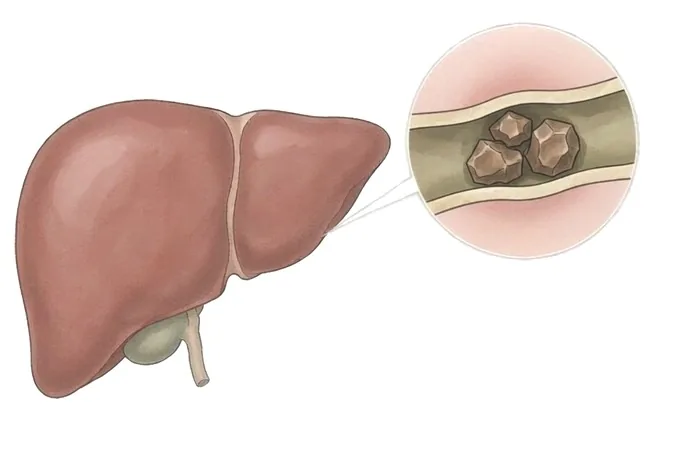
Stones can sit in the gallbladder, or they can sit in the bile ducts themselves — the channels that carry bile from the liver to the bowel. Stones in the ducts cause a different disease from gallbladder stones: they block drainage, they infect easily, and the infection can move fast. A stone inside the ducts within the liver — hepatolithiasis — is the form seen far more often in East and Southeast Asia than in the West, and it tends to be a long-term disease of repeated attacks rather than a single event. The good news is that almost every part of it is treatable, provided the disease is recognised for what it is and mapped before anything is done.
-
Fever + jaundice + pain
the classic triad of acute cholangitis — same-day assessment
-
Drainage before stone removal
during an infection the duct is drained first, and cleared after
-
MRCP maps the whole tree
every duct, stone and stricture, before any treatment is planned
-
48 hrs
from complete records to a personalized MDT plan
What bile duct stones are — and why they differ from gallbladder stones
The liver makes bile, the bile drains through a branching tree of bile ducts into the bowel, and the gallbladder hangs off that tree as a storage bag. Most people know about gallstones, which sit in the bag. A separate and in some ways more serious disease is stones that sit in the ducts themselves: in the main channel outside the liver (the common bile duct), or in the branches inside the liver — which is what hepatolithiasis means. This page covers the duct disease in both locations.
The difference matters because of what ducts cannot do. The gallbladder tolerates holding stones; it is a bag designed to store bile, and many people carry gallbladder stones for life without trouble. A duct is a channel, not a store: a stone in it blocks drainage, and blocked bile backs up into the liver (which shows as jaundice) and infects easily (which shows as cholangitis — fever with shaking chills). A blocked, infected duct is an active medical problem, not something to watch.
Where the stones come from separates two groups. Stones in the main duct either form there or migrate from the gallbladder, and the treatment follows the origin. Stones inside the liver usually form there, on a combination of poor drainage, narrowed ducts and repeated infection, and they are often accompanied by duct strictures and by segments of liver that have slowly scarred around them. In East and Southeast Asia this intrahepatic form is far more common than in Western countries, and historically it has been bound to parasitic infection carried by raw freshwater fish — a link that still shows up today.
The shape of the disease follows from all this. Main-duct stones often declare themselves with one dramatic episode — pain, jaundice, infection — and can often be cleared in one or two endoscopic sessions. Intrahepatic stones tend to be a long-term disease: repeated attacks over years, ducts that narrow and re-block, and liver segments that slowly fail. That is why the diagnostic map (the section on how the ducts are imaged) and the choice between endoscopy and surgery matter so much, and why follow-up after treatment is not optional.
Types by location: inside the liver, in the main duct, and in between
Location decides almost everything about bile duct stones — how they present, what treatment reaches them, and what the long-term plan looks like. The four groups below cover the disease.
| Type | Where the stone sits | Typical story and treatment |
|---|---|---|
| Gallbladder stones that migrated | In the main duct outside the liver, having slipped down from the gallbladder | Often one episode of blockage or pancreatitis; cleared endoscopically, and the gallbladder is usually removed to stop it happening again |
| Primary main-duct stones | Formed in the main duct itself, usually where bile stagnates or infection persists | Endoscopic clearance is the core treatment, with attention to why bile stagnates — a narrowed duct or a duct that does not empty |
| Intrahepatic stones (hepatolithiasis) | In the duct branches inside the liver, often on one side, with strictures and dilated ducts around them | A long-term disease of repeated attacks; treated with a combination of endoscopy, percutaneous routes and sometimes removal of the affected liver segment |
| Recurrent pyogenic cholangitis pattern | Intrahepatic and main-duct stones together, with repeated bacterial infection, historically linked to liver flukes from raw freshwater fish | The full combination: drainage during attacks, stone clearance, treatment of strictures, and long-term surveillance |
Two words used in reports are worth unpacking. Choledocholithiasis means stones in the common bile duct — the main channel — regardless of where they formed. Hepatolithiasis means stones in the ducts inside the liver. A person can have both at once, and when the intrahepatic disease has been present for years the picture often includes duct strictures, segments of liver that have thinned or scarred, and a history of attacks that were treated with antibiotics each time but never cured. That combined picture is what the older literature calls recurrent pyogenic cholangitis, and it is the reason the intrahepatic disease is treated as a liver disease rather than a plumbing problem.
Two axes of severity: the acute attack and the years of disease
Bile duct stone disease is graded along two axes that are kept separate because they answer different questions. The first is the acute attack: how sick is the person today, and how fast must the duct be drained? This is what the severity grades for acute cholangitis describe. The second is the chronic disease: what have the years done to the ducts and to the liver? The first decides the pace of treatment; the second decides its extent.
| Grade of the acute attack | What it looks like | What it demands |
|---|---|---|
| Mild | Fever and pain, blood tests abnormal, organs still working normally | Antibiotics and prompt drainage; many of these patients are candidates for stone removal at the same endoscopic session once the plan is clear |
| Moderate | Signs that the attack is affecting the blood, kidneys or clotting, or a longer course | Drainage without delay, a longer course of treatment, and stone clearance deferred to a second sitting once the infection has settled |
| Severe | Organ dysfunction — confusion, falling blood pressure, failing kidneys — the emergency picture of severe cholangitis | Intensive care and drainage as the urgent priority, by whatever route is fastest; definitive stone treatment waits until the patient has stabilised |
The chronic axis is assessed on the MRCP and on liver imaging: how many ducts hold stones, where the strictures are, whether ducts above a blockage are permanently dilated, and whether any segment of liver has scarred, thinned or stopped working. A single stone in a healthy main duct and a liver half-taken by stone disease with strictures are both "bile duct stones", but they are not the same condition and they do not get the same plan. The combination of both axes — how bad today is, and how much disease the years have built — is what the treatment plan is written against.
How the ducts are mapped, and what each test answers
The tests follow a sequence, and each one answers a different question. The rule that matters most: the ducts are mapped before they are treated. Clearing stones through a duct tree nobody has imaged is how stones get left behind and strictures get missed.
| Test | What it answers |
|---|---|
| Liver function tests | Whether the pattern points to blockage — bilirubin and the cholestatic enzymes rising together — and how inflamed the system is |
| Abdominal ultrasound | The first image: dilated ducts, often the stones themselves, and the gallbladder |
| MRCP | The map: every duct, every stone, every stricture, without any intervention — the scan the whole plan is written from |
| CT | Detail the MRI cannot give: large stone loads, changes in liver segments, and complications of long-standing disease |
| ERCP | Both an examination and a treatment — used when drainage or stone removal is itself the next step, not as a first look |
Two assessments sit outside the duct tree but decide the plan. The first is the liver itself: where intrahepatic disease has been present for years, the segments that hold stones may have scarred and stopped contributing, and the segments that remain have to carry the load — which is measured with scans and blood tests before anyone talks about removing liver tissue. The second is the gallbladder: if it holds stones too, it is part of the same disease, and leaving it in place invites the next migration.
Why patients travel to China for bile duct stone treatment
Bile duct stone treatment rewards exactly the things a large East Asian hepatobiliary centre is built around. The intrahepatic form of the disease — the form that needs the most judgement — is far more common here than in the West, which means the teams who treat it see it continuously rather than occasionally, and the full toolkit for it is in daily use rather than on paper.
- The full toolkit in one place. Emergency drainage, endoscopic stone removal, duct-opening by balloon and by mechanical lithotripsy, cholangioscopy through the working channel of the scope, percutaneous routes through the liver, and the surgical options from duct exploration to segment resection and drainage reconstruction — the decision about which combination applies is a genuine choice here, not a forced one by what happens to be available.
- Imaging-first discipline. MRCP maps the whole duct tree before treatment, and the plan is written against the map rather than decided scope-in-hand.
- Speed where it counts. Fever with jaundice is treated as a same-day problem; drainage is arranged around the clock, and definitive treatment follows once the infection has settled.
- 48-hour written MDT plan from complete records — images themselves, not just reports — so that the second opinion and the treatment plan can be formed before travel.
- Cost at roughly 30%–50% of Europe or the US, with more than 30 international insurers on direct billing.










