


Hepatic Hemangioma Overview
Hepatic Hemangioma Overview
Of all the things a liver scan can find unexpectedly, a hemangioma is the most common — and the most reassuring once it is understood. A hemangioma is a tangle of blood vessels, present from development, growing slowly if it grows at all, and it does not turn into cancer. The word tumour in a report describes a mass, not a malignancy, and for the great majority of patients the correct treatment is a firm diagnosis followed by nothing more than observation — or, for larger lesions, a period of imaging follow-up to confirm stability. The real work in this disease is diagnostic: confirming with confidence what the lesion is, so that nobody is operated on for something that never needed surgery.
-
Not cancer, and never becomes it
the word tumour means a mass; hemangiomas do not turn malignant
-
Diagnosed by imaging, not biopsy
contrast CT or MRI shows a near-signature pattern
-
Observation is the default
follow-up scans confirm stability; most need nothing further
-
48 hrs
from complete records to a personalized MDT plan
What a hepatic hemangioma is — vessels, not cancer
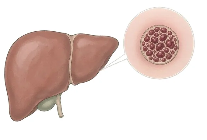
A hemangioma is a benign tumour made of blood vessels — literally a tangled, oversized cluster of the small vessels that ordinary tissue is full of. In the liver it is usually the cavernous type: blood-filled spaces separated by thin walls, like a sponge soaked in blood. It is the most common benign tumour of the liver, and one of the most common incidental findings on any abdominal scan.
Three facts do most of the work in this disease, and they are worth stating plainly. First, a hemangioma is not cancer and does not become cancer — the transformation everyone fears has no place in this lesion’s biology. Second, it is present from development: this is something the liver was built with, not something that grew there later. Third, it grows slowly if it grows at all; most lesions never change measurably across decades.
Why, then, does it deserve a cluster of pages? Because of what happens in the months after the word appears in a report. A lesion in the liver raises the question of cancer, and the diagnosis has to be earned with confidence before that question is retired. Most of the anxiety patients bring to this clinic comes not from the hemangioma but from an earlier stage at which someone said tumour without saying benign, or said watch it without saying why. The cure for that is a diagnosis made properly: the right scans, read by people who see liver lesions every day.
The shape of the management follows. Where the imaging is characteristic, the diagnosis is secure and most patients need nothing further. Where the lesion is larger, a period of follow-up imaging confirms stability. Where the lesion causes symptoms — the minority — or the images leave genuine doubt, treatment is planned: and the options, from removing the lesion to blocking its blood supply, are chosen deliberately rather than urgently. What almost never happens is the emergency the patient feared on reading the report.
Types: cavernous, and the variations that matter
Hemangiomas of the liver are classified by their structure and by their number, and the classification is simple because the disease is simple:
| Type | What it is | What it means |
|---|---|---|
| Cavernous hemangioma | Blood-filled spaces separated by thin walls — the sponge-like structure, and by far the commonest form | The standard lesion this guide describes; diagnosed on imaging, observed or treated as the size and symptoms dictate |
| Small capillary-type lesions | Tiny, dense vessel clusters, often only a few millimetres | Almost always incidental; confirmed by their appearance and left alone |
| Multiple hemangiomas | Several lesions at once (hemangiomatosis) | Still benign; each lesion is read on its own imaging features, and follow-up covers all of them |
| Giant hemangioma | A lesion large enough to occupy much of the liver — a size descriptor, not a different disease | Assessed for symptoms, compression and growth; these are the lesions in which treatment, rarely, is discussed |
One distinction belongs here because it is the other lesion a report may mention: a hemangioma is not the same as focal nodular hyperplasia or hepatic adenoma, the other common benign liver lesions. All three are benign, but they behave differently — adenomas, unlike hemangiomas, are influenced by hormones and can require treatment, and telling the three apart on imaging is exactly the diagnostic work this centre does daily. A report that says only liver lesion, without saying which, has not yet answered the question.
Size and symptoms: two axes that decide the plan
Hemangiomas are graded along two axes that are read together. The first is size — the common convention divides lesions into small, large and giant, though exact cut-offs vary slightly between centres. The second is what the lesion does: nothing, pressure symptoms, or the rare event. Size alone never dictates treatment; the combination does.
| Category | Typical picture | What it asks for |
|---|---|---|
| Small lesion, no symptoms | Found incidentally; characteristic imaging; nothing to feel | A firm diagnosis and, in most cases, no follow-up beyond the first confirmation |
| Large lesion, no symptoms | Still silent, but large enough that stability is worth confirming | Follow-up imaging at set intervals; treatment only if it grows or starts to matter |
| Giant lesion | Occupies a substantial part of the liver; may press on neighbouring organs | Full assessment — symptoms, compression, growth over time — with treatment discussed on its merits |
| Symptomatic lesion, any size | Fullness, pain, early satiety attributable to the lesion, or rapid growth | The situation in which treatment is genuinely considered — planned, not urgent |
The second axis deserves one honest paragraph. Attributing symptoms to a hemangioma is done carefully, because the upper abdomen is a crowded place and gallbladders, stomachs and acid all produce symptoms of their own. A lesion that is simply present while a patient has dyspepsia is not thereby the cause of it. This is why the assessment asks not only what the lesion looks like but whether the story fits — and it is why so many consultations here end with treating something other than the hemangioma, or with the quiet conclusion that the hemangioma is an innocent bystander.
How the diagnosis is confirmed — and why no biopsy
Hemangiomas are diagnosed from images, and the sequence is built around one idea: the scans already know the answer, so the biopsy is skipped on purpose.
| Step | What it answers |
|---|---|
| Ultrasound | Where most are first seen — a bright, well-defined spot; often the end of the story for typical small lesions |
| Contrast-enhanced CT | The filling pattern: hemangiomas enhance from the edge inward, a recognisable signature |
| MRI with contrast | The most distinctive examination — a near-signature appearance that settles doubtful cases |
| Blood tests | Liver function — usually normal — and, in very large lesions, counts that screen for rare consumption syndromes |
| Repeat imaging | Stability over time — the test that closes the loop for larger lesions |
The deliberate absence from this table is biopsy, and the reason is worth knowing. A needle into a mass of blood vessels adds bleeding risk for almost no informational gain — the scans answer the question — and a biopsy of a hemangioma can be actively misleading, producing bloody, uninterpretable material. The clinical rule is that a lesion with a characteristic imaging pattern does not need tissue, and a lesion that does not have one needs better imaging, not a needle. This is one of the clearest cases in medicine where doing less is doing more, and it is the first thing to confirm if someone has recommended a biopsy to you.
Why patients travel to China for a liver-lesion opinion
A liver lesion needs two things in combination: readers who see enough of them to be certain, and a centre where the rare minority that needs treatment can be treated without needing to travel again. Both sit together here.
- High-volume reading. Liver lesions are among the most common incidental findings in imaging, and the hepatobiliary teams here characterise them daily — the difference between probable and confident is volume.
- The full toolkit in one place. Ultrasound, contrast CT, dedicated liver MRI — and, for the small minority who need it, liver surgeons who remove hemangiomas while sparing the rest of the liver, with interventional radiology alongside.
- Restraint as a discipline. The correct answer for most patients is no treatment, and saying so confidently — with the reasoning written down — is the service.
- 48-hour written MDT plan from complete records, so the second opinion and the plan are formed before travel.
- Cost at roughly 30%–50% of Europe or the US, with more than 30 international insurers working with us on direct billing.










