


Esophageal Cancer Overview
Esophageal Cancer Overview
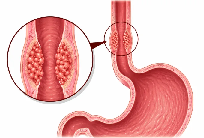
The oesophagus is a muscular tube with no outer coat and a dense network of lymph channels in its wall, so a cancer arising from its lining spreads early. The type of cancer and its depth decide whether it can be removed through an endoscope, by an operation, or by chemoradiotherapy.
-
~50%
of the world's oesophageal cancer cases are diagnosed in China
-
Two types
squamous cell carcinoma and adenocarcinoma — different causes, different geography
-
HER2
amplified in a proportion of adenocarcinomas, which makes targeted treatment an option
-
48 hrs
from complete records to a personalized MDT plan
What esophageal cancer is
The oesophagus is a muscular tube about 25 centimetres long that carries food from the throat to the stomach, passing through the neck, the chest and the diaphragm on the way. Its wall has a mucosal lining, a submucosal layer containing mucus glands and a dense lymphatic network, a muscle layer, and an outer adventitia of loose connective tissue — but no serosa, which is the slick outer membrane that encloses most of the gut.
That anatomical difference explains a great deal about this cancer. Without a serosa, there is no barrier to keep a tumour from reaching neighbouring structures — the trachea, the aorta, the pleura, the pericardium — and the lymph channels in the submucosa run lengthwise along the tube, so tumour cells can travel well beyond the visible edge of the lesion before they reach a node. This is why early oesophageal cancer spreads to nodes earlier and further than its size would suggest, and why the lymph nodes are removed as part of the operation rather than sampled.
Two other features shape the disease worldwide. It has two histological types with different causes — squamous cell carcinoma and adenocarcinoma — and they have different geographic distributions, which means the same organ hosts two diseases that are prevented and screened for in different ways. And it has a detectable precursor stage: high-grade dysplasia in squamous epithelium and Barrett's oesophagus with dysplasia in glandular epithelium can both be treated before an invasive cancer develops, which is unusual among gastrointestinal cancers and is the basis of screening in high-incidence regions.
The main types
Oesophageal cancer is classified twice over — by what the cells look like and by where in the tube they arise — and both classifications influence treatment:
- Squamous cell carcinoma — arises from the flat cells lining the upper and middle oesophagus. It is the dominant type in China, Central Asia and Iran, is linked to tobacco, alcohol and very hot drinks, and it accounts for roughly half of the world's cases being diagnosed in China. It responds to radiotherapy, which is why chemoradiotherapy plays such a large part in its treatment.
- Adenocarcinoma — arises from glandular cells near the junction of the oesophagus and stomach, almost always on a background of Barrett's oesophagus, in which chronic reflux has replaced the normal squamous lining with glandular mucosa. It dominates in Western countries and is linked to reflux and central obesity rather than to hot drinks.
- Barrett's oesophagus and dysplasia — not a cancer, but the treatable precursor of adenocarcinoma, and the reason surveillance programmes exist. High-grade dysplasia is treated rather than watched.
- By location — cervical, upper thoracic, middle thoracic, lower thoracic and gastro-oesophageal junction. The location decides the operation, the radiotherapy field, and in the cervical oesophagus whether surgery is offered at all.
- Rare types — small cell carcinoma, neuroendocrine tumours, melanoma and lymphoma of the oesophagus. Each behaves differently and is treated on its own terms rather than as squamous carcinoma.
The practical consequence is that a treatment plan cannot be built on "oesophageal cancer" alone. Squamous carcinoma of the middle third and adenocarcinoma of the lower third differ in their risk factors, their preferred chemotherapy, their radiotherapy fields and the operation used — which is why the pathology report, the location and the depth are all needed before anything is decided.
What each stage means
Staging is done with endoscopic ultrasound for depth and nodes, and with CT and PET-CT for the chest, abdomen and distant spread. The AJCC system then combines the depth of invasion (T), the nodes (N) and distant spread (M) into stages:
| Stage | In plain terms | How it is usually approached |
|---|---|---|
| 0 | High-grade dysplasia or carcinoma in situ — abnormal cells confined to the surface lining | Endoscopic removal or ablation; no operation needed |
| I | Tumour reaches only the mucosa or the submucosa, with no involved nodes | Endoscopic resection for mucosal disease; oesophagectomy for submucosal disease |
| II | Tumour reaches the muscle layer with no nodes, or a mucosal tumour with one or two involved nodes | Surgery, usually preceded by chemoradiotherapy |
| III | Tumour reaches the outer adventitia, or several regional nodes are involved | Chemoradiotherapy first, then surgery; or definitive chemoradiotherapy |
| IVA | Tumour involves a structure that cannot be removed — the aorta, the trachea or the vertebral column — or many nodes are involved | Definitive chemoradiotherapy, with surgery in selected cases after a response |
| IVB | Distant spread — most often to the liver, lungs, bones or distant lymph nodes | Systemic treatment, with stenting or radiotherapy for swallowing |
Two boundaries inside this table carry most of the decisions. The first is the muscularis mucosae: a tumour confined above it can be removed with a scope, while one that has entered the submucosa has access to the lymph channels and usually needs an operation. The second is the T4a and T4b distinction: tumour touching the pleura, pericardium or diaphragm can still be removed with those structures, while tumour invading the aorta, the trachea or a vertebral body cannot, and the plan changes from surgery to chemoradiotherapy.
Because both boundaries are decided by depth rather than by length, the staging tests are not interchangeable. CT shows how far the tumour has spread but not how deep it has gone; endoscopic ultrasound is the test that measures depth, and it is what separates a lesion that can be treated endoscopically from one that needs an operation.
The markers that guide treatment
Oesophageal cancer has fewer drug targets than lung or breast cancer, but a short list changes treatment for definable groups of patients, and testing is done on the biopsy:
| Marker | What it is | How it changes management |
|---|---|---|
| HER2 (ERBB2) | Over-expression or amplification of a growth-factor receptor, found in a proportion of adenocarcinomas | Supports adding trastuzumab to first-line chemotherapy in advanced disease, and is being tested in earlier stages |
| PD-L1 | An immune checkpoint protein reported as a combined positive score from the biopsy | Supports a PD-1 inhibitor in advanced disease and, after chemoradiotherapy with residual disease, in the adjuvant setting |
| Mismatch repair and MSI status | A rare pattern in which DNA repair errors accumulate | Makes immune checkpoint inhibitors substantially more likely to work, and is worth testing for whenever immunotherapy is being considered |
| TP53 | The commonest altered gene in squamous cell carcinoma | Not directly targetable, but it supports the diagnosis and is part of the molecular picture reported alongside the type and grade |
The practical point is that the tissue from the diagnostic endoscopy is what makes all of this possible, which is one reason a biopsy is taken at the first examination rather than at a later one. Asking for HER2, PD-L1 and mismatch repair status at the time of diagnosis avoids a second procedure later.
Why patients come to China for esophageal cancer care
Oesophageal cancer is unusual in that the country with the largest number of cases is also where the greatest concentration of treatment experience sits — and it is one of the cancers where that experience is measurable in the operation itself.
- Volume in oesophageal resection — oesophagectomy is among the most demanding operations in gastrointestinal surgery, involving three compartments and two joins, and the outcomes at a centre performing it weekly are not those of a centre performing it occasionally.
- Minimally invasive oesophagectomy — thoracoscopic and laparoscopic techniques that avoid a large chest incision and a laparotomy, with the lymph node dissection performed through the same keyhole approach.
- Three-field lymph node dissection — removal of nodes in the neck, chest and upper abdomen, which matters because the lymphatic drainage of the oesophagus runs lengthwise and skip metastases are common.
- Endoscopic treatment of early lesions — submucosal dissection and mucosal resection for high-grade dysplasia and mucosal cancer, so that the earliest lesions never need an operation.
- Chemoradiotherapy with modern delivery — intensity-modulated and image-guided radiotherapy, with concurrent chemotherapy, given either before surgery or as definitive treatment.
- Screening experience that transfers — because China runs endoscopic screening in high-incidence regions, the ability to recognise and treat early lesions is part of routine practice rather than an occasional skill.
Two practical consequences follow. The correct treatment may be an endoscopic procedure, an operation or chemoradiotherapy, and which of the three applies depends on the depth of invasion — so the endoscopy report and the endoscopic ultrasound matter as much as the CT. And because chemoradiotherapy may be given before surgery rather than after it, the sequence should be settled before travel. Send complete records and the written assessment is returned within 48 hours of the file being complete.










