


Endometriosis Overview
Endometriosis Overview
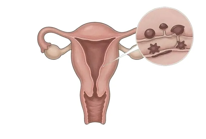
Endometriosis is the name given to tissue that resembles the lining of the womb growing where the lining of the womb does not belong. It is found on the peritoneum that lines the pelvis, on an ovary, in the tissue between the vagina and the rectum, on the bowel, on the bladder, or inside a caesarean scar. It is not cancer and it is not an infection. What makes it matter is that it behaves like the tissue it resembles: it thickens and breaks down with the cycle, and the blood it produces has nowhere to go. The result is inflammation, then bleeding into surrounding tissue, then scarring and adhesions that tether organs to one another. That is why the pain is cyclical at the beginning, and why it can become continuous as the disease and the scarring progress. It is also why the amount of disease found does not predict how much it hurts. Two facts run through everything that follows. Pain and fertility are separate lines, and improving one does not automatically improve the other. And the diagnosis is usually delayed for years, which means that waiting for certainty rather than treating on the clinical picture is itself a decision, with its own cost.
-
The pain does not match the disease found
a few small implants can cause pain that stops someone working, and extensive disease can be found in someone with few symptoms
-
A normal scan excludes almost nothing
the commonest form sits on the peritoneal surface and is beyond the resolution of any imaging
-
Treatment can begin before the diagnosis is confirmed
where the picture is typical, waiting for a laparoscopy is a choice with its own cost rather than a neutral step
-
Pain and fertility are two separate lines
one set of decisions is made on the pain history and another on the fertility assessment, and they do not answer each other
What endometriosis is, and what the name gets wrong
It is a condition defined by where tissue is, not by an infection or a growth that has to be removed:
- The tissue is like the lining of the womb, and it is in the wrong place. It behaves as the lining behaves — thickening and breaking down with the hormonal cycle — but there is no outlet. The blood it produces is released into the surrounding tissue instead, and that is the origin of everything that follows: inflammation first, then scarring, then adhesions that pull organs together and hold them there.
- Pain and disease are not proportional. A few small implants on the peritoneum can produce pain that stops someone working, and extensive disease can be found in someone who came for an unrelated reason and has few symptoms. This is not a matter of pain tolerance; it is that the symptoms come from inflammation and irritation of nerve-rich surfaces rather than from the volume of tissue present.
- It is usually cyclical at the start and can become continuous. Early on the pain tracks the period, because the tissue responds to the cycle that drives it. As scarring and adhesions accumulate, the pain loses that pattern and becomes a background that is present most days — a change worth reporting precisely because it marks a shift in the disease.
- It is commoner than the name suggests, and it is not rare. A great many women live with it for years under the heading of normal period pain before anyone describes it as a condition. The problem is not that it is obscure; it is that its symptoms are usual enough to be dismissed and its commonest form is invisible on every scan.
- It is not the same condition as adenomyosis. Adenomyosis is tissue like the lining of the womb present within the muscle of the womb itself. It shares the cyclical pain and the heavy periods, it is coded separately, and it is treated differently — which matters because the two can occur together and one is easily mistaken for the other.
What the word does not mean. It does not mean cancer, and it does not become cancer. It does not mean the pain is psychological, although living with it for years has psychological consequences that deserve treatment in their own right. It does not mean the disease is caused by anything a woman did, ate or failed to do. It does not mean a pregnancy is impossible, and it does not mean that a pregnancy will cure it. What it means is that tissue is present in the wrong place, that the amount of it and the amount of pain are different questions, and that there are two lines of treatment to consider separately.
The five places it is found, and what each one changes
Where the disease sits matters more than how much of it there is, because the site determines the symptoms and the treatment:
| Where it is found | What follows from it |
|---|---|
| On the peritoneum lining the pelvis | The commonest form and the one that no scan can see. It is reached at laparoscopy, and it is the form most often behind years of pain that was called unexplained |
| On or inside an ovary | An endometrioma, sometimes called a chocolate cyst, which ultrasound does identify. It changes the plan because removing it also removes some normal ovarian tissue, so the reserve is discussed before surgery |
| Deeply, between the vagina and the rectum, or into the bowel | Pain on sex, on opening the bowels and on passing urine around the period. Mapping it and treating it involves the disciplines that would be needed if those organs are opened |
| On the bladder or the ureter | Cyclical urinary symptoms, and in some cases a kidney that is silently affected, which is a reason to look even when the pain is modest |
| In a caesarean or perineal scar | Cyclical pain and swelling in a scar that is otherwise healed. It is uncommon, it is easy to recognise once considered, and it is treated by excision |
Why the site matters more than the volume. Two women with the same quantity of disease can have entirely different problems: one has pain limited to the period because the disease is superficial, another has pain on sex and on the bowel because a small amount of disease sits deep in a place that hurts with every movement. A plan written from the volume of disease rather than from its position treats the wrong thing.
What changes the plan, and how the pace is set
What changes the plan is not how extensive the disease sounds on a report but what it is doing to the person and what they want next:
| What changes the plan | What it changes |
|---|---|
| How much the pain is affecting work, sleep and relationships | It decides whether the first step is medical treatment for pain, surgery, or both, and it is the measure the plan is reviewed against |
| Whether a pregnancy is wanted now, later, or not at all | It decides which of the two lines comes first, because some treatments control pain while preventing pregnancy and some do the opposite |
| Whether the bowel, bladder or ureter is involved | It changes who performs surgery and what is discussed beforehand, and it raises the threshold for operating rather than lowering it |
| How many operations have already been performed | Repeat surgery is considered differently, because each operation carries its own scar and the ovarian reserve does not return |
| Whether the pain is still cyclical or has become continuous | A change from cyclical to continuous suggests scarring rather than active disease, which shifts what is likely to help |
Why pace is the practical question. Couples who ask how serious it is usually mean how quickly they should move and what to agree to first. Those answers come from how the pain is affecting daily life, what the fertility line needs, and how many operations have already been done — which is why both lines are discussed in the same consultation rather than one after the other.
What the assessment is looking for
The assessment does three things: it takes the history by pattern, it uses imaging for what it can show, and it looks for what is being missed:
| What is assessed | What it establishes |
|---|---|
| The pain history, taken by pattern | When in the cycle the pain occurs, whether it comes with sex, with the bowel or with passing urine, and whether it has become continuous, which together are the most informative part of the assessment |
| A pelvic examination | Whether the pelvis is fixed and tender, whether there is a nodule behind the cervix, and whether an ovary is enlarged, all of which change what is suspected |
| A transvaginal ultrasound | An ovarian endometrioma and many deep lesions, when performed by an operator looking for them — and, when negative, the important fact that a normal scan excludes almost nothing |
| An MRI where deep disease is suspected | The map of the disease, used to plan surgery and to decide which disciplines need to be present rather than to establish the diagnosis |
| Tests for the other explanations | Whether the pain is from pelvic inflammatory disease, irritable bowel syndrome, bladder pain syndrome or something else, because each is treated differently and some occur alongside endometriosis |
| The fertility line, assessed separately | Ovarian reserve, ovulation and tubal patency where relevant, because the fertility question is answered by its own examination rather than inferred from the pain |
Why the assessment is deliberately two-tracked. Almost every item on this list changes what is done next. What the assessment is not is a search for certainty before anything can begin: where the picture is typical, treatment for pain can start on the clinical diagnosis while the fertility line is assessed in parallel, and laparoscopy is reserved for the point at which it will change what happens.
Why patients come to China for this assessment
This is a condition whose problems are spread across several specialties, and travelling for it is usually about having them answered together:
- The history is taken by pattern rather than by severity. When the pain occurs, what else it comes with, and whether it has changed are recorded as the diagnostic information they are, rather than reduced to a severity score.
- Imaging is used for what it can show. Ultrasound is performed by someone looking specifically for deep disease, MRI is used to map it before surgery, and a negative scan is read as an absence of evidence rather than as evidence of absence.
- Surgery is planned with the ovarian reserve in mind. Where an endometrioma is to be removed, the effect on the reserve is discussed before the operation rather than discovered afterwards, and repeat surgery is treated as a decision with a cost.
- Pain and fertility are assessed as two lines in one visit. The pain plan and the fertility assessment are separate sets of decisions, and both are made before the plan is written rather than one deferred until the other is settled.
- The plan and the records are prepared to travel. Imaging is issued with the dates and the technique used, and the written plan names which line is being treated, what is being watched, and when the next review is, so that care continued at home continues from a document rather than from memory.
What this does not promise. A completed assessment does not guarantee that the pain will be controlled, that a pregnancy will follow, or that the disease will not recur — recurrence is the expected behaviour of the condition, not a failure of treatment. Saying which parts can be acted on and which cannot is part of the assessment rather than a caveat added afterwards.










