


Endometrial Cancer Overview
Endometrial Cancer Overview
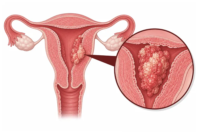
The endometrium is the lining of the uterus, rebuilt and shed with every cycle under the control of estrogen and progesterone. When estrogen acts for years without the opposing hormone, the lining can grow abnormally and, in some women, become cancerous. The disease is the commonest gynaecological cancer in high-income countries — but it is also one that announces itself early, because the same lining that turns malignant bleeds when it breaks down.
-
No. 1
the commonest gynaecological cancer in high-income countries (WHO/IARC)
-
4 classes
POLE-mutant, MMR-deficient, p53-abnormal and NSMP — molecular classes that now shape the plan
-
~5%
of cases arise within Lynch syndrome, a hereditary DNA repair condition (IARC)
-
48 hrs
from complete records to a personalized MDT plan
What endometrial cancer is
The uterus is a muscular organ lined by the endometrium, a layer of tissue that thickens under estrogen each cycle and is shed when progesterone withdraws. Cancer begins when cells in this lining accumulate changes that let them grow without the cycle's discipline. Because the lining sits in a hollow organ and bleeds whenever it breaks down, the disease usually gives an early, unmistakable signal — abnormal bleeding — long before it has spread anywhere.
Most tumours follow years of estrogen acting without opposing progesterone ("unopposed estrogen"): after menopause, adipose tissue becomes the main source of estrogen, which is why weight is the strongest modifiable risk factor, and the same logic explains risks tied to never being pregnant, late menopause and estrogen-only hormone therapy. A smaller group arises outside this hormonal pathway — serous and clear-cell carcinomas, and tumours in women with Lynch syndrome — and behaves more aggressively.
The disease spreads in an orderly way: first into the muscle wall of the uterus (the myometrium), then to the cervix, the ovaries and tubes, and the pelvic and para-aortic lymph nodes, and only late to distant organs. Because the uterus is removed in almost every case, surgery is the backbone of treatment, and the depth of muscle invasion plus the molecular class decide what happens after it.
The main types
Endometrial cancer is classified by the cell it arises from and the behaviour that follows, and the type feeds directly into staging and treatment:
- Endometrioid carcinoma — the commonest type, driven by unopposed estrogen, graded G1 to G3 by how much gland-like structure it keeps, and usually found early. Low-grade tumours (G1–G2) behave indolently; high-grade (G3) behaves more like the aggressive types.
- Serous carcinoma — an aggressive type that does not depend on estrogen, can arise in a thin, atrophic lining, and spreads even when the main tumour is small. It is treated on the same lines as ovarian serous cancer.
- Clear-cell carcinoma — uncommon, also aggressive, and likewise managed as high-risk disease regardless of how early it appears.
- Carcinosarcoma — a mixed tumour containing both carcinoma and sarcoma elements, managed as high-risk with chemotherapy in the plan.
- Precancer (endometrial hyperplasia with atypia) — a thickened, disordered lining that is not yet cancer; hysterectomy or progestin therapy at this stage prevents cancer from developing.
Pathology reports also carry the two findings that modern staging counts: lymph-vascular space invasion and the molecular markers described below. Both travel with the specimen from the first biopsy onward, which is why the biopsy report — not just the ultrasound — belongs in the records sent for a second opinion.
What each stage means
Staging uses the FIGO 2023 system, which keeps the anatomical framework — tumour confined to the uterus, spread beyond it, distant disease — and now amends each stage by the molecular class. The anatomical stages relevant to decisions are:
| Stage | In plain terms | How it is usually approached |
|---|---|---|
| IA | Tumour confined to the lining, or invading less than half the muscle wall | Surgery alone in most cases; no radiotherapy beyond follow-up for low-risk disease |
| IB | Invading half or more of the muscle wall | Surgery; post-operative radiotherapy decided by grade and molecular class |
| II | Tumour has grown into the cervical stroma | Surgery; adjuvant radiotherapy commonly considered |
| III | Spread to the ovary or tube, the outer surface of the uterus, or pelvic / para-aortic nodes | Surgery followed by chemotherapy, often with radiotherapy |
| IVA | Invasion of bladder or bowel lining | Chemoradiotherapy or systemic treatment |
| IVB | Distant spread, including peritoneum, lung or liver | Systemic therapy — immunotherapy where the molecular class allows |
Two features of FIGO 2023 matter in practice. First, the molecular class moves the stage: a POLE-mutated tumour with high-risk anatomy is reclassified to a better prognosis category, while a p53-abnormal tumour confined to the lining is moved upward, because its behaviour resembles serous cancer. Second, node status is explicit — involved pelvic nodes place disease in stage III, which is why sentinel node mapping is part of the operation for anything beyond the lowest risk.
Because the amendments apply to tumours of all grades, the molecular tests are requested on the original biopsy wherever possible — they can be ready before surgery, and the surgical plan plus the adjuvant discussion follow from them.
The markers that guide treatment
Endometrial cancer was among the first cancers where molecular classification entered routine staging. A panel of four tests — POLE sequencing, mismatch-repair immunohistochemistry and p53 staining — assigns every tumour to one of four classes with distinct behaviour:
| Class | What it is | How it changes management |
|---|---|---|
| POLE-mutated | A mutation in the DNA proof-reading gene, producing enormous numbers of mutations | The best-prognosis class; adjuvant treatment is often de-escalated even at high anatomical stage |
| MMR-deficient | Loss of mismatch-repair proteins, found in roughly a quarter to a third of cases | Makes the tumour responsive to immune checkpoint inhibitors in advanced or recurrent disease |
| p53-abnormal | Abnormal p53 staining, marking tumours that behave like serous cancer | The highest-risk class; adjuvant therapy is escalated and treatment follows the serous-cancer lines |
| NSMP | No specific molecular profile — the largest class | Management follows grade, stage and additional markers such as L1CAM; a substantial subgroup has an intermediate outlook |
The practical point: ask whether POLE, MMR and p53 have been tested, and on which specimen. Where they have not, the tests can often be run on the original biopsy tissue — no new procedure needed — and the result may change both the operation planned and what follows it. The same MMR result that refines prognosis is what unlocks immunotherapy later, so it is worth having on file from the start.
Why patients come to China for endometrial cancer care
Endometrial cancer surgery rewards volume and precision — a high-volume gynaecological oncology service that does the whole pathway, from molecular pathology to minimally invasive surgery to sentinel node mapping, under one roof.
- Minimally invasive surgery as standard — total hysterectomy with removal of tubes and ovaries done laparoscopically or robotically for apparent early-stage disease, with faster recovery and fewer wound complications than open surgery.
- Sentinel node mapping — dye- and tracer-guided removal of the first draining nodes, which spares most patients a full nodal dissection and the leg-swelling that can follow it.
- Molecular pathology in-house — POLE, mismatch-repair and p53 testing reported on the biopsy, so the molecular class informs the surgical and adjuvant plan rather than arriving after it.
- Immunotherapy access — PD-1 inhibitors for mismatch-repair-deficient disease, and pembrolizumab with lenvatinib where markers do not allow it, arranged within the network.
- Fertility-preserving programmes — progestin therapy with structured hysteroscopic surveillance for young women with low-grade disease who wish to keep the uterus.
- Coordinated follow-up — international patients keep imaging and examination schedules across time zones through the network's international service.
Two practical consequences follow. The choice of surgery — and whether nodes are sampled — depends on MRI findings and the biopsy's grade and molecular class, so all of those should be sent, not just the ultrasound. And because adjuvant treatment is decided by the molecular class, records from the original pathology matter as much as the imaging. Send complete records and the written assessment is returned within 48 hours of the file being complete.










