


Diabetic Retinopathy Overview
Diabetic Retinopathy Overview
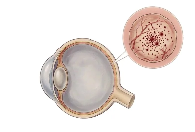
Diabetic retinopathy is damage to the small blood vessels of the retina caused by diabetes, and it is a leading cause of vision loss in adults of working age. Its defining feature is silence: through the stages in which treatment works best there are no symptoms at all — no pain, no blurring, nothing that would send a patient to an eye doctor — which is why the disease is found by examination rather than by how the eye feels. The damage follows a recognisable order. Years of high blood sugar weaken the walls of the small vessels; they bulge, leak fluid and lipid into the retina, and then begin to close, leaving patches of retina short of oxygen. In some eyes the deprived retina responds by growing new vessels, which are fragile in exactly the way that matters: they bleed into the eye and later contract, pulling on the retina. Alongside the vessel disease sits a second, separate problem — swelling of the macula, the small central area that reads and recognises faces — which blurs the centre of the vision and can occur at almost any stage. The course of the disease is dominated by how long diabetes has been present, but it is not fixed by it: glucose, blood pressure and lipid control slow it, and laser, injections or surgery applied at the right stage protect the sight that remains. What no treatment does is restore retinal tissue that has already been lost, which is why the honest strategy is to find the disease while the patient can still feel nothing.
-
It gives no warning
at the stages when treatment works best there are no symptoms at all — screening finds the disease, not symptoms
-
Two problems, one name
vessel damage that can progress to fragile new vessels, and swelling of the macula — different findings, different treatments
-
Duration dominates, control changes the course
years with diabetes matter most, but glucose and blood pressure control measurably slow the damage
-
48 hrs
from complete records to a written assessment plan
What diabetic retinopathy is, and why silence is the problem
Diabetic retinopathy is damage to the small blood vessels of the retina — the light-sensitive layer at the back of the eye — caused by diabetes. It is one of the commonest complications of long-standing diabetes and a leading cause of vision loss in adults of working age. Its defining feature is silence: at the stages where treatment is most effective, the eye feels entirely normal, there is no pain, and the vision may be unchanged. The disease is therefore found by examination rather than by complaint, and everything about managing it follows from that fact.
The mechanism is slow and orderly. High blood sugar over years weakens the walls of the small vessels that feed the retina; they bulge into small pouches, leak fluid and lipid into the retinal tissue, and then begin to close, leaving patches of retina without oxygen. In some eyes the starved retina responds by growing new vessels — which are not replacements but fragile, misdirected tissue that bleeds into the eye and later contracts, pulling on the retina it sits on.
- The damage and the swelling are two different problems. The vessel disease itself is staged from mild to proliferative, and swelling of the macula — diabetic macular edema — is a separate finding that can appear at almost any stage. They threaten sight in different ways and are treated with different tools, so an assessment always answers for both.
- Vision is lost late, not early. The centre of the retina keeps working while the disease is still treatable, which is why waiting for blurring before being examined means waiting past the point where treatment is simplest. The symptoms that eventually appear — blurring, floaters, a bleed — belong to stages that screening was meant to prevent.
- The course is changeable, and part of the change is the patient’s. Duration of diabetes is the strongest single factor, and it cannot be altered. What can be altered is the environment the vessels live in: glucose, blood pressure and lipids. Good control does not make retinopathy impossible, but it slows it measurably — which is why control and screening travel together rather than substituting for each other.
What treatment can and cannot do. Treatment can stop or slow the vessel disease, seal leaks around the macula, make new vessels shrink and disappear, and remove blood or membranes that threaten the retina. What it cannot do is restore retinal tissue that has already been lost. The aim, stated plainly, is to protect the sight the patient still has — and the sight worth protecting is greatest at the stages when the patient notices nothing.
The stages diabetic retinopathy passes through
Retinopathy is graded by what the retina shows, because the grade decides what happens next — reassurance and review, local treatment, urgent laser, or surgery. Four stages of vessel disease are recognised, and one further finding runs alongside them.
| What it is called | What is happening | How it behaves |
|---|---|---|
| Mild non-proliferative retinopathy | Small pouches (microaneurysms) appear in the vessel walls — the earliest visible change | No symptoms. Managed with continued control and regular review, which is how progression is caught early |
| Moderate non-proliferative retinopathy | More vessels are affected; some leak fluid and lipid, some begin to close | Still usually silent. Review intervals shorten, and the macula is watched specifically for swelling |
| Severe non-proliferative retinopathy | Widespread vessel closure leaves large areas of retina short of oxygen | The stage at which treatment is discussed before new vessels grow — the window that screening exists to find |
| Proliferative retinopathy | New vessels grow on the retina or elsewhere in the eye, fragile and prone to bleed and contract | The stage of bleeding, scar tissue and traction on the retina. Treated promptly with scatter laser, injections or surgery |
| Diabetic macular edema | Fluid accumulates in the macula, the small central area that does detailed vision | Can occur at almost any stage, including mild. The commonest cause of moderate vision loss in diabetes, and treated with injections or laser |
Why the staging is not academic. Each stage has a different decision attached to it, and the difference between stages is information that only the examination can supply. A patient with severe non-proliferative disease feels exactly the same as a patient with mild disease; what separates them is the state of their vessels, and the treatment that is worth starting in one and not yet in the other.
Where macular edema sits in the picture. It is listed alongside the stages rather than within them because it does not follow them in sequence. A patient can have mild vessel disease with significant swelling, or proliferative disease with a dry macula. Both findings are assessed at every visit, and the more urgent of the two sets the plan.
What makes retinopathy more or less urgent
Within any stage, a short list of findings decides how quickly things must happen. All of them are established in the same assessment.
| Feature | Why it changes the plan |
|---|---|
| Whether the macula is swollen | Swelling involving the centre threatens reading vision directly and is usually treated now rather than watched; its extent is measured, not guessed |
| Whether new vessels are present | Proliferative disease is treated promptly because the complications it is heading for — bleeding, traction, a detached retina — are harder to treat than the vessels themselves |
| How far the vessel disease has gone | Severe non-proliferative disease sits just before proliferation; at this stage treatment is discussed in advance rather than after the new vessels arrive |
| The state of the other eye | Retinopathy is usually bilateral but rarely equal; each eye is graded separately, and the worse eye sets the tempo while the better eye is protected by the same discipline |
| The general diabetes picture | Kidney disease, blood pressure and glucose control are read together with the retina, because they move in the same direction and affect both the disease and the safety of any treatment |
What urgent means in this disease. It rarely means the same day, as it does in some eye emergencies. It means that findings which predict rapid deterioration are acted on in weeks rather than left to the next annual review — and that the patient knows which category they are in, because the plan is written down rather than implied.
How a diabetic retina is assessed
The assessment answers four questions in order — is there damage, how far has it gone, is the macula swollen, what should be done — and each step exists to make the next decision precise.
| Step | The question it answers |
|---|---|
| Visual acuity in each eye | Is the centre still working? A fall in the reading acuity is the first clue that the macula is involved, and it is the number treatment is judged against |
| Dilated fundus examination | What do the vessels, the macula and the periphery show? This is the step on which the stage itself is based, and both eyes are examined at every visit |
| Optical coherence tomography | Is the macula swollen, and by how much? OCT measures the fluid and gives a thickness number that monitoring and treatment can be compared against |
| Fluorescein angiography | Where exactly are vessels leaking, and where have they closed? Used when that map would change the plan — before laser, or to explain vision loss that the examination does not account for |
| Review of the blood picture | What is the HbA1c, the blood pressure, the kidney function? The retina is read together with the diabetes, because the same process is running in vessels everywhere |
Why the order matters. The acuity and the dilated examination establish what is happening; the imaging quantifies it; the blood results explain the environment it is happening in. An assessment that produced a stage without the macular measurement, or a treatment plan without the blood picture, would be incomplete in the ways that lead to the wrong interval between visits.
Why patients travel to China for diabetic retinopathy care
Diabetic retinopathy is a disease of schedules rather than single events: the outcome is set by how reliably the retina is examined, measured and treated at the right intervals. What an international pathway offers is that the whole assessment can happen inside one visit instead of being spread across weeks of referrals.
Examination and imaging are completed the same day. Dilated examination, macular imaging, angiography where it is needed and a review of the blood picture are all available in one facility, so a patient who arrives with no symptoms and a decade of diabetes leaves the same day with a stage for each eye, a measured macula and a written plan.
Treatment can begin within the same visit where it is indicated. Injections and laser are performed without a waiting period, and if surgery is needed the decision, the preparation and the operation are arranged inside one admission — which matters in a disease where the interval between finding and treating is part of the treatment.
Both eyes, and the whole diabetic picture, are handled together. Each eye is graded separately, the blood results are reviewed with the retina, and where cataract or other eye disease coexists it is built into the same plan rather than referred elsewhere.
The follow-up schedule is written for someone going home. Which eye is examined when, what the macular thickness should be doing between visits, which symptoms require immediate attention and which measurements the next treating team should inherit are provided in English; the international desk coordinates with the patient, with any insurer or assistance company, and with the treating team at home.










