


Craniocerebral Trauma Overview
Craniocerebral Trauma Overview
Craniocerebral trauma is injury to the brain caused by an external force — a fall, a collision, a blow, or the acceleration of the head itself. The most useful thing to understand about it is that it is two injuries rather than one. The primary injury happens at the moment of impact and nothing done afterwards can undo it. The secondary injury happens over the following hours and days — low oxygen, low blood pressure, raised pressure inside the skull, fever, seizures, and a collection of blood that was not found and goes on enlarging — and it is the half that medicine can actually change. This guide follows the order families need: what the first signs are and why they are graded on response rather than on appearance, the patterns of injury and which ones need surgery, how severity is graded and what recovery really looks like, and the two presentations that arrive by getting better and then worse.
-
48 hours
written opinion once complete records arrive
-
One team
neurosurgery, neurocritical care, neuropsychology and rehabilitation planning together
-
Emergency imaging
non-contrast CT reported for blood, fracture and shift, with repeat scanning on any fall in consciousness
-
124+
countries and regions our international patients come from
What craniocerebral trauma is — and what it is not
Craniocerebral trauma is injury to the brain caused by an external mechanical force. It covers the whole range from a concussion that resolves in a fortnight to an injury requiring surgery within the hour, and the range matters less than understanding one thing about it: it is two injuries, not one.
The primary injury happens at the moment of impact. The brain is bruised against the skull at the point of contact and again on the opposite side, the long connections between nerve cells are stretched and torn, blood vessels are torn, and the skull itself may fracture. Nothing done in the hours afterwards can undo any of that. This is the part families find hardest to accept, and it is worth saying plainly rather than implying that prompt treatment will erase it.
The secondary injury happens in the hours and days afterwards, and it is the half that medicine actually operates on. Oxygen that does not reach the brain. Blood pressure that is too low to perfuse it. Pressure inside the skull that rises and pushes the brain against its own openings. Fever. High blood sugar. Seizures that consume what reserve is left. And a collection of blood that was small on the first scan and goes on enlarging. Every one of these has a treatment, every one of them runs on a clock, and together they account for more of the final outcome than the force of the original impact does.
Two consequences follow, and they explain most of the management. First, the first minutes matter most: getting oxygen in and blood pressure up is the intervention associated with the largest difference in outcome, and it happens before anyone reaches a scanner. Second, observation is a treatment, not a passive wait. The purpose of watching someone in a unit that can escalate is to catch the deterioration that secondary injury announces before it has finished happening.
What it is not: it is not a stroke, though a head injury can be followed by one and the two are sometimes hard to separate in the first hours. It is not a degenerative disease. It is not caused by anything the person did wrong, and in the case of a fall in an older adult, it is very often caused by something else that has not yet been looked for — a medicine, a rhythm, a blood pressure, an unsteady gait. How force becomes injury is set out on the causes page.
The patterns of injury, and which one needs surgery
The same force produces different injuries depending on where it lands, how fast the head was moving, and whether it stopped suddenly or rotated. Naming the pattern is practical, because it decides who goes to theatre within the hour and who is watched.
| Pattern | What it is and how it presents | What to look for alongside | What the result changes |
|---|---|---|---|
| Concussion | A disturbance of how nerve cells work rather than a structural change: confusion, a memory gap, headache, dizziness, nausea | The scan is normal by definition; the diagnosis is clinical, from the mechanism plus the symptoms | No surgery. Management is a graded return to activity rather than prolonged rest |
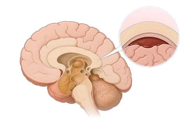
| Extradural haematoma | Bleeding between the skull and the brain's outer covering, usually from a torn artery after a fracture | The lucid interval — briefly clear, then rapid deterioration; a lens-shaped collection on the scan | Urgent surgical evacuation. The pattern where an hour changes everything, and the reason a normal early scan does not end observation |
| Acute subdural haematoma | Bleeding between the brain's coverings, usually from torn bridging veins, often with injury beneath | Rapid deterioration after impact; a crescent-shaped collection; more common in older adults and with anticoagulation | Usually surgical; the underlying brain injury, not the collection, sets the ceiling on recovery |
| Chronic subdural collection | The same bleed, accumulating slowly over weeks after a fall that seemed trivial | An older adult; confusion, unsteadiness, a changed personality or failing memory rather than a headache | Often drained through a small opening with rapid improvement. The most treatable pattern and the most often missed |
| Contusion | Bruising of the brain itself, typically at the front and undersides of the frontal and temporal lobes | Can enlarge over the first day or two and behave like a collection; anticoagulation makes this likelier | Usually watched with repeat imaging; surgery if it enlarges or raises pressure |
| Diffuse axonal injury | Widespread tearing of the long connections between nerve cells, from acceleration and rotation rather than a blow | Prolonged unconsciousness from the moment of impact, with a scan that can look surprisingly mild | No surgical target. Care is prevention of secondary injury plus rehabilitation, and the scan understates it |
| Skull fracture | A break in the skull, open or closed, depressed or linear | Raises the probability of a collection beneath it; a base-of-skull fracture can leak cerebrospinal fluid | The fracture itself often needs no treatment; what matters is what lies beneath it |
| Penetrating injury | An object that has entered the skull; the damage follows its track rather than the force | Infection, seizure and vascular risk along the track | Surgical debridement, antibiotics and seizure prevention, in a centre that does this regularly |
Two points follow. More than one pattern is usually present — a person with a subdural collection also has a contusion and, quite often, an element of diffuse injury, and the one that needs the theatre first is the one that is compressing or shifting the brain. And the pattern can change over the first day: a contusion that looked small on the first scan can be a surgical collection on the second. How that change is caught is set out on the diagnosis page.
How severity is graded, and what recovery looks like
Severity in this disease is graded on what the person does, not on how the injury looks. A person shouting is not necessarily less badly injured than a person lying still. The Glasgow Coma Scale records three things — whether the eyes open, whether speech makes sense, and whether movement obeys a command — and it is recorded as three component scores rather than as one number, because the number hides the detail that the decision depends on.
| Grade or phase | What it looks like | What it means for care | What it does not tell you |
|---|---|---|---|
| Mild | Alert, or momentarily confused; scan usually normal; symptoms settle over days to weeks | Discharge with a written plan and someone who knows what to watch for; graded return, not prolonged rest | Not that the brain was unaffected. A concussion is invisible to a scanner, and a minority have persisting symptoms |
| Moderate | Drowsy or confused, with a scan that often shows something; the level of consciousness is watched hour to hour | Admission to a unit that can escalate; repeat imaging on any fall in consciousness; early rehabilitation referral | It does not predict the outcome. Moderate injuries move both ways over the first day |
| Severe | Not obeying commands, sometimes not opening the eyes or speaking; the scan usually shows a collection or diffuse injury | Neurocritical care from the first hour: airway, oxygen, blood pressure, and pressure measured rather than guessed | A low score in the first minutes is not a verdict. Sedation, intubation and low blood pressure all move it |
| The first forty-eight hours | The window in which secondary injury happens: a collection enlarges, pressure rises, a seizure occurs | Repeat imaging on deterioration rather than on a schedule, to catch a change while it is still reversible | A stable first scan is not a stable course. Most of what goes wrong here goes wrong after it |
| The recovery weeks to months | Improvement that is uneven: awareness and basic function first, then mobility, then speech, then speed of thinking and mood | Rehabilitation throughout, reviewed rather than restarted at each plateau; decisions on the trend, not the day | A plateau is not the end of improvement, which continues for months and often years after a severe injury |
| Persistent symptoms after a mild injury | Headache, dizziness, poor sleep, irritability and difficulty with sustained concentration beyond the expected weeks | An active programme rather than more rest: graded activity, sleep treatment, headache management, return in steps | It is not permanent damage and not untreatable. Prolonged rest is one of the things that keeps it going |
| What follows afterwards | Post-traumatic headache, epilepsy, sleep disturbance, mood change, and after severe injury, memory and impulse-control difficulty | Treated on its own terms; each has a treatment, and none improves by being waited out | Their presence is not a new injury. They are recognised consequences, managed rather than merely monitored |
What nobody can promise. Not a cure, and not a return to exactly the previous level. What can be said honestly: after a mild injury most people recover fully over weeks; after a severe injury recovery is measured in months to years and is often uneven, with the residual difficulties most often falling in the areas of speed of thinking, memory, stamina, mood, sleep and impulse control. The two things within medicine's control that decide the most are oxygen and blood pressure in the first minutes, and pressure inside the skull afterwards.
How the assessment is assembled
The assessment runs in an order set by the clock rather than by convenience: airway, breathing and circulation first, then the response, then the scan, then the decision. Nothing about it is complicated; what makes it work is doing the parts in that order and repeating the parts that move.
| Step | What it involves | What it establishes | What it does not rule out |
|---|---|---|---|
| Airway, breathing, circulation | Oxygen given, blood pressure supported, the neck protected until the spine is cleared | Whether the two secondary injuries with the largest effect on outcome are being prevented now | Nothing else matters until this is right. It precedes the scan rather than competing with it |
| The mechanism | What force, what height, what speed, who saw it, and whether the head stopped suddenly or rotated | Which patterns are likely, whether the neck needs imaging, and whether a bystander's account exists | A trivial-sounding mechanism does not exclude serious injury, especially in an older adult on anticoagulation |
| The Glasgow Coma Scale | Eye opening, speech and movement, recorded as three components and repeated to the same standard | Severity, and more usefully the direction of travel between one recording and the next | A single number hides detail, and is moved by sedation, intubation, low blood pressure and alcohol |
| Pupils | Size and reactivity, recorded and repeated | Whether the brain is being compressed and on which side — a pupil that stops reacting is a surgical sign | A normal pupil does not exclude a collection. It is the change in a pupil that carries the information |
| Emergency non-contrast CT | Reported for blood, fracture, shift and pressure, by someone looking for all four | Whether there is a collection that needs the theatre, and whether the brain is being pushed | A normal scan excludes neither a concussion, nor diffuse axonal injury, nor a collection that has yet to enlarge |
| Repeat CT on deterioration | The same scan again when the level of consciousness falls rather than rises, or a pupil changes | Whether a collection has enlarged — the commonest cause of a deterioration absent on arrival | Repeated on a schedule rather than on a change, it finds less and exposes more. The trigger is the examination |
| MRI at 3.0T where indicated | Imaging where symptoms persist with a normal CT, or where the extent of injury needs characterising | Diffuse injury, small contusions, and the patterns computed tomography cannot resolve | Not a screening test after every head injury, and it does not itself change acute management |
| EEG where indicated | Routine recording, extended with video where a seizure or unexplained fluctuation in consciousness is in question | Whether an event was a seizure, and whether there are seizures not visible from the bedside | A normal recording excludes neither. Subclinical seizures occur after severe injury and are worth looking for |
The principle underneath all of it: the examination is the monitor, and the scan is what you do when the monitor changes. Most of what goes wrong in this disease goes wrong after the first scan, which is why the diagnosis page is organised around what to repeat and when rather than around what to order once.
Why patients travel to China for a head injury assessment
Patients come for three reasons: an injury that was assessed and discharged but has not settled; a scan that was reported as normal while symptoms persist; or a question about whether something that is being watched at home should still be watched. The value here is not a different operation — it is a team that runs imaging, neurosurgery, neurocritical care and rehabilitation in one pathway, and that says plainly what the scan shows, what the trajectory is, and what would change the answer.
What the pathway looks like. Neurosurgery, neurocritical care, neuroradiology, neuropsychology and rehabilitation plan together. Emergency non-contrast CT with repeat scanning on any change in consciousness, 3.0T MRI, dual-source CT, PET-CT, 3D DSA and long-term video EEG sit inside the same programme, as do pressure monitoring and the operating theatre. A written opinion is returned within 48 hours of complete records arriving. Fully treated costs typically run at 30–50% of comparable care in Europe or the United States, more than 30 commercial insurers are billed directly, and our international patients come from 124 countries and regions.
What a records-only opinion can and cannot do. It can say whether the imaging was adequate and whether it was repeated when it should have been, whether the pattern on the scan is one that needs intervention now or one that is watched, whether there is a delayed complication the follow-up has not looked for, and whether the rehabilitation is matched to what the person is actually struggling with. It cannot examine the person, repeat a scan, or measure the pressure inside the skull — and those are sometimes exactly what the case turns on. We say so rather than implying otherwise, and where a visit would add nothing we say that too, before any payment is discussed.
What must be said about timing. An acute head injury is a local emergency and is treated where it happens; nobody should travel during the first hours of a significant injury, and no assessment here justifies it. The right moment to travel is afterwards — when the question is why symptoms have not settled, whether a delayed complication has been missed, or what the rehabilitation should now be. Send the mechanism and the timeline first: what force, what date, what the person was like before, and what has changed since. That is the part of the file no hospital can reconstruct later.










