


Coronary Heart Disease Overview
Coronary Heart Disease Overview
Coronary heart disease is not one disease but a spectrum. At one end sits a narrowed artery that only complains when the heart is asked to work harder — predictable, manageable, and controllable for years with medicine. At the other end sits a ruptured plaque and an artery closing in minutes, where the amount of heart muscle that survives is decided by how quickly the artery is reopened. Between the two ends sit all the questions this guide answers: which symptoms matter, which test answers which question, and when medicine, a stent or a bypass is the right answer rather than a matter of preference.
-
24/7
Emergency cardiac intervention cover at both flagship hospitals
-
170
Specialised cardiovascular beds across the two flagship hospitals
-
5
National certifications at the Foshan hospital — the first in its city
-
124+
Countries and regions our international patients come from
What coronary heart disease is — a pipe problem with a clock
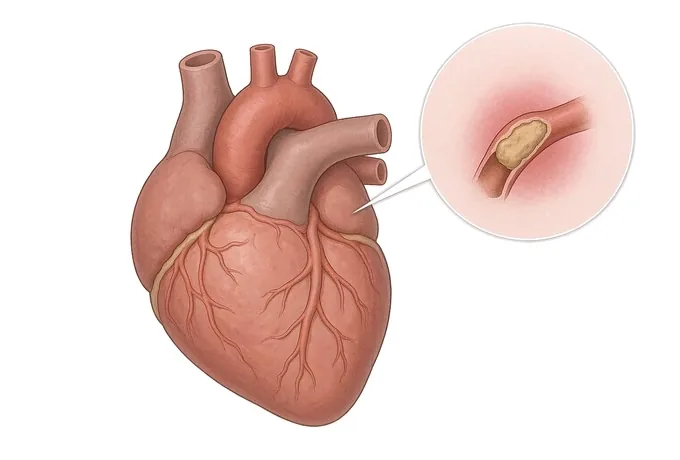
The heart is a muscle, and muscle needs its own blood supply. That supply arrives through three small arteries — the coronary arteries — that run on the surface of the heart and are between one and four millimetres wide. Over decades, cholesterol carried in the blood can accumulate in their walls and form plaque: a mixture of lipid, fibrous tissue and calcium that thickens the wall and narrows the channel. That process is atherosclerosis, and when it happens in the coronary arteries the condition is called coronary heart disease or coronary artery disease — two names for the same thing.
What the narrowing does depends entirely on how quickly it develops. A plaque that grows slowly over years gives the heart time to adapt: the artery narrows, and the first complaint arrives only when the demand outstrips the supply — climbing a hill, carrying luggage, walking against a cold wind. That is angina, and although it is unpleasant it is not itself an emergency. A plaque that ruptures is a different event: the torn surface triggers a clot that can close the artery in minutes, and the muscle it supplies begins to die. That is an acute coronary syndrome — the medical term that covers unstable angina and heart attack — and in that event the clock is the treatment: the sooner the artery is reopened, the more muscle survives.
Three consequences organise the rest of this guide. First, the absence of symptoms does not exclude the disease: a plaque can narrow an artery by half without ever causing pain, which is why risk factors and imaging, not symptoms alone, drive the diagnosis in many patients. Second, the artery that is blocked matters as much as how blocked it is — a 70% narrowing in a small side branch and the same percentage in the left main artery, which supplies most of the heart, are entirely different problems. Third, treatment is chosen from a comparison, not a reflex: medicine, stenting and bypass each have situations in which they are clearly the best answer, and the diagnostic work exists to find out which situation the patient is in.
One distinction belongs here because patients arrive carrying it: coronary heart disease is not heart failure and not a valve problem. All three can coexist — a long-narrowed heart can weaken, and rheumatic valve disease is its own guide elsewhere — but each has its own page, because their treatments differ. This guide is about the arteries.
The spectrum: stable disease and the acute syndromes
Coronary disease is classified first by how it is behaving right now, because that decides whether the next hours are routine or an emergency — and then by where the blockages sit, because that decides which treatments are candidates. The two classifications are independent: a patient with one blocked artery can present as an emergency, and a patient with three blocked arteries can be entirely stable.
| Category | What is happening | What it means for treatment |
|---|---|---|
| Chronic coronary syndrome (stable angina) | Plaques that have been present for years, narrowing the artery predictably under exertion; symptoms arrive with effort and leave with rest | Not an emergency. Medicine is the foundation; revascularisation — stent or bypass — is added when the anatomy and the amount of ischaemia justify it |
| Unstable angina | A plaque has become active: pain at rest, new-onset pain, or pain climbing in severity, without heart-muscle damage on the blood test | An emergency in slow motion — admitted, treated with medicine immediately, and usually investigated within days |
| NSTEMI | The same ruptured-plaque process, with a rise in troponin showing that some muscle has been damaged | An emergency: risk-scored, and the artery is usually opened during the same admission |
| STEMI | A complete blockage of a major artery, visible on the electrocardiogram; muscle is dying minute by minute | The clock event: the artery is reopened as fast as possible, around the clock — the situation behind the 24/7 emergency cover at our two flagship hospitals |
| Silent ischaemia | Narrowings that starve the muscle without ever causing pain — most often in diabetes and in older patients | Found on testing rather than by symptoms; treated on the same anatomy-and-ischaemia logic as painful disease |
The second classification is anatomical, and it is the one that decides between a stent and a bypass. The positions that matter: the left main artery, which supplies about three quarters of the heart and is the highest-stakes position; the proximal left anterior descending artery, the long vessel down the front of the heart; multivessel disease, where two or three arteries are narrowed; and chronic total occlusion — an artery that has been blocked for months or years, whose reopening is a procedural specialty of its own. Where each blockage sits, how many there are, and whether the anatomy suits wires or grafts — that comparison is made on the angiogram and is the subject of the treatment page.
Two axes: the anatomy of the blockages, and what the muscle still does
Coronary disease has no stage numbers in the way cancer does, and patients sometimes find that disorienting: the angiogram report contains percentages and names of arteries rather than a single stage. In practice the severity is read on two axes together, and they are kept separate on purpose. The first is anatomical — where the blockages are and how complex they are. The second is functional — how much the symptoms limit the patient, and whether the muscle is genuinely short of blood.
| Axis | What is measured | Why it matters |
|---|---|---|
| Anatomy: position | Which artery and which segment — left main, proximal LAD, other major vessels, side branches; the vessel supplying the rest of the heart through collaterals | Position, more than percentage, decides how dangerous a narrowing is and which treatment suits it |
| Anatomy: complexity | How many vessels are diseased; whether a blockage is a chronic total occlusion; calcification and tortuosity that make wires or grafts harder | Complexity is the raw material of the stent-versus-bypass comparison, and of the procedural plan |
| Function: symptom load | The CCS angina grade, from I (ordinary activity does not provoke angina) to IV (any activity, or angina at rest) | The grade describes what the disease costs the patient today, and it is the yardstick against which any treatment's benefit is judged |
| Function: ischaemia | Evidence that the muscle is short of blood — exercise testing, stress imaging, or a pressure-wire measurement taken at angiography | A moderate-looking narrowing without ischaemia is treated with medicine; the same narrowing with a large territory at risk changes the plan |
| Function: muscle state | Echocardiographic pumping function (ejection fraction), and whether parts of the muscle are scarred from a previous event or merely stunned and recoverable | Weakened muscle shifts decisions: it raises the stakes of complete revascularisation and favours surgery in multivessel disease |
Three things are worth saying before treatment is discussed. First, a percentage is not a verdict: a 60% narrowing that the pressure wire shows is not restricting flow needs no stent, and that single measurement has redirected many patients away from procedures they did not need. Second, the two axes must be read together: a modest-looking narrowing in the left main artery outranks an impressive-looking one in a small branch, and the report that treats every 70% as equal has not been read carefully. Third, severity describes the arteries, not the person: the same anatomy in a fit seventy-year-old and in a patient with weak kidneys, severe lung disease or diabetes leads to different advice, because the durability of each option has to be weighed against the life it will be living in.
How the diagnosis is made, and what each test answers
Each test answers a different question, and the order matters: the wrong order wastes either time — which is muscle in an acute event — or a catheter that the patient did not need.
| Test | The question it answers | What it cannot do |
|---|---|---|
| Electrocardiogram (ECG) | Is an artery closing now? Is the rhythm stable? Has a silent infarction left its trace? | A normal ECG does not exclude coronary disease — most stable plaques leave it untouched |
| High-sensitivity troponin | Has muscle been damaged — the blood test that separates a heart attack from unstable angina | It says nothing about the anatomy; a normal troponin does not mean the arteries are clean |
| Echocardiogram | How well the muscle pumps, whether a wall moves abnormally, and whether the valves are involved | It sees the consequences of narrowings, not the narrowings themselves |
| Coronary CT angiography | Are the arteries involved at all — a scan that maps the wall and excludes significant disease without a catheter | Heavily calcified arteries are hard to grade, and treatment cannot usually be delivered through the same sitting |
| Invasive coronary angiography | The definitive map — every artery, every narrowing, the collaterals; and the doorway through which stents are delivered | It shows the channel, not the wall, and by itself it does not prove that a moderate narrowing is starving the muscle |
| Pressure-wire measurement (FFR / iFR) | Whether an intermediate narrowing genuinely restricts flow — measured during the angiogram, before the decision to stent is made | It adds procedural time and is not needed for every lesion — it is spent where the answer would change the plan |
Two points of practice follow. The first is that the pathway is chosen by the question: exclude disease in a lower-risk patient and the CT scan is the efficient answer; treat a high-risk or acute patient and the angiogram is, because treatment can follow in the same sitting. The second is that the diagnosis is not complete until the report answers four things: how the arteries are affected and where, whether the concerning narrowings are genuinely ischaemic, what the pumping function is, and what the risk score says about urgency. A report that says only "coronary artery disease" leaves every treatment decision unmade.
Why patients travel to China for coronary treatment
Coronary treatment is decided from images and risk scores, and both travel well. The classification systems are international, the devices are the same drug-eluting stents used worldwide, and the operations — angioplasty and bypass — are standardised. What separates providers is the span of the service: whether the emergency, the complex intervention and the surgery sit under one roof, and how quickly a visiting patient moves from records to a decision.
Our cardiovascular service spans two flagship hospitals — Fosun Hospital Foshan and Fosun Hospital Guangzhou — with nearly 170 specialised cardiovascular beds, modern coronary care units and advanced catheterisation laboratories. Emergency cover runs around the clock. The Foshan hospital holds national certifications in five areas — chest pain, atrial fibrillation, heart failure, cardiac rehabilitation and hypertension — the first hospital in its city to do so, and the Guangzhou hospital obtained national chest pain centre certification in 2025. Beyond standard stenting, the teams treat the complex anatomy that other centres refer on: left main and multivessel disease, chronic total occlusions, and bypass surgery with on-site surgical backup supported by intra-aortic balloon pumping when it is needed.
For a visiting patient, records reviewed by a cardiology team receive a written opinion within about 48 hours of complete records arriving — the angiogram films or discs themselves, not only the report, are what the review needs. Complete treatment in China typically runs at 30–50% of comparable care in Europe or the United States, we work with more than 30 commercial insurers for direct billing, and our international patients have come from 124 countries and regions. Cardiac rehabilitation — the part of coronary care that decides whether the result holds — is delivered through a nationally certified programme rather than left to a leaflet.










