


Cervical Cancer Overview
Cervical Cancer Overview
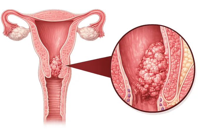
The cervix is the lower part of the uterus, and almost every cervical cancer begins after an HPV infection that the body failed to clear. Because the infection is preventable and the precancer is visible, this is among the most preventable of cancers — and when it does develop, the stage decides whether treatment is a cone biopsy, an operation, or radiotherapy with chemotherapy.
-
~99%
of cervical cancers are linked to a persistent HPV infection (WHO)
-
HPV 16 / 18
two viral types account for roughly 70% of cases — and are covered by every vaccine
-
FIGO 2018
the staging system for this cancer now explicitly includes imaging findings
-
48 hrs
from complete records to a personalized MDT plan
What cervical cancer is
The cervix is the narrow lower part of the uterus that opens into the vagina, and its surface carries two kinds of lining: squamous cells on the outer portion facing the vagina, and glandular cells lining the canal that leads up into the uterus. The junction between the two — the transformation zone — is where almost all cervical cancers begin, and it is precisely the area a screening test samples.
The disease develops slowly in most cases. A high-risk HPV infection, acquired through sexual contact, is cleared by the immune system within a year or two in the great majority of people. When it persists instead, the viral genes disturb the way the lining cells mature, first producing cervical intraepithelial neoplasia (CIN) — a precancer confined to the surface — and, over years, an invasive cancer that has grown through the basement membrane into the underlying tissue. That long interval between infection, precancer and cancer is the reason screening works here better than for almost any other cancer.
It also spreads in a predictable pattern, which matters for treatment: first into the tissue beside the cervix (the parametrium) and the upper vagina, then to the lymph nodes of the pelvis and later those along the aorta, and only in advanced disease to distant organs. Surgery removes the cervix and the parametrium together for early disease, while radiotherapy plus chemotherapy covers the same territory without an operation for locally advanced disease — which is why stage, more than anything else, decides the plan.
The main types
Cervical cancer is classified by the cell it arises from, and the type influences both how it is detected and how it behaves:
- Squamous cell carcinoma — arises from the flat cells on the outer surface, is the commonest type worldwide, and is strongly linked to HPV 16. It is the type most readily caught by conventional cytology.
- Adenocarcinoma — arises from the glandular cells of the cervical canal. Its share has been rising everywhere, it sits higher in the canal where a smear samples less reliably, and HPV testing catches it better than cytology does.
- Adenosquamous and rare types — tumours with both components, and uncommon varieties such as neuroendocrine or small cell carcinoma. Each is treated on its own protocol rather than as an ordinary cervical cancer.
- Precancer (CIN / AIS) — cervical intraepithelial neoplasia on the surface and adenocarcinoma in situ in the canal are not cancer, but treating them is what prevents cancer from ever developing.
The type is reported on every pathology specimen, and it carries practical consequences: adenocarcinoma argues for HPV-based screening and careful assessment of the canal, rare types can change the chemotherapy used, and both influence how follow-up is arranged after treatment.
What each stage means
Staging uses the FIGO 2018 system, which — unlike its predecessors — explicitly incorporates MRI and PET-CT findings, and even pathology from removed nodes. The stages relevant to treatment decisions are:
| Stage | In plain terms | How it is usually approached |
|---|---|---|
| IA1 | Microscopic invasion no deeper than 3 mm, found on a cone or a biopsy | Cone biopsy alone in most cases; no hysterectomy required |
| IB1 | Tumour 2 cm or smaller, confined to the cervix | Radical hysterectomy — or radical trachelectomy where fertility matters |
| IB2–IB3 | Tumour between 2 and 4 cm, or larger than 4 cm, still confined to the cervix | Surgery for IB2; chemoradiotherapy and brachytherapy for IB3 |
| IIB | Tumour has grown into the parametrium, the tissue beside the cervix | Definitive chemoradiotherapy with brachytherapy |
| IIIC | Pelvic nodes involved (IIIC1) or para-aortic nodes involved (IIIC2) | Chemoradiotherapy with an extended field; surgical staging may precede it |
| IVA / IVB | Invasion of bladder or rectal lining, or distant spread to lung, liver, bone | Systemic treatment, with radiotherapy for bleeding or symptom control |
Two boundaries inside this table carry most of the decisions. The first is tumour size at 2 and 4 centimetres: up to 2 cm, fertility-sparing surgery is an option; beyond 4 cm, most guidelines move to chemoradiotherapy instead of surgery. The second is the parametrium: once tumour reaches it, an operation is generally replaced by radiotherapy with concurrent chemotherapy, because the radiotherapy field covers the same tissue more completely than surgery can.
Because FIGO 2018 counts involved nodes as stage IIIC even when the tumour itself is small, the imaging report directly changes the treatment category — which is why MRI and PET-CT are done before the plan is set, not after.
The markers that guide treatment
Cervical cancer is driven by viral oncogenes rather than by a wide range of targetable mutations, but a short list of markers determines eligibility for immunotherapy and is tested on the biopsy:
| Marker | What it is | How it changes management |
|---|---|---|
| PD-L1 | An immune checkpoint protein reported as a combined positive score (CPS) from the biopsy | A CPS of 1 or more supports adding pembrolizumab to chemotherapy, with or without bevacizumab, in advanced disease |
| Mismatch repair / MSI status | A rare pattern in which DNA repair errors accumulate | Makes a PD-1 inhibitor likely to work regardless of PD-L1, and is worth requesting whenever immunotherapy is considered |
| Tumour mutational burden (TMB-H) | An unusually high number of mutations per megabase | An alternative route to pembrolizumab where PD-L1 is negative but the tumour is mutation-rich |
| NTRK fusions | Very uncommon gene fusions reported in occasional cases | Qualify for TRK-inhibitor treatment when no standard option remains |
The practical point: request PD-L1 and mismatch repair testing at the time of diagnosis rather than when advanced disease appears, because the original biopsy is usually the best tissue available and a later re-biopsy can be difficult to arrange.
Why patients come to China for cervical cancer care
Cervical cancer care rewards exactly the things a high-volume national network provides — brachytherapy quality, surgical volume, and screening infrastructure that recognises precancer on sight.
- Brachytherapy as a core skill — internal radiotherapy delivered into the cervix is what makes chemoradiotherapy complete for locally advanced disease, and centres that perform it routinely plan it with MRI guidance.
- Nerve-sparing radical surgery — radical hysterectomy that removes the parametrium while preserving bladder, bowel and, in selected cases, sexual function, done by open or minimally invasive approach.
- Fertility-sparing operations — radical trachelectomy and large-cone procedures for tumours of 2 cm or less, performed by teams who also manage the pregnancies that follow.
- Immunotherapy access — PD-1 inhibitors combined with chemotherapy and bevacizumab for advanced disease, with the molecular testing arranged in-house.
- Screening and colposcopy depth — China runs large-scale cervical screening, so the ability to read colposcopy accurately and to treat CIN before it becomes cancer is routine practice.
- Coordinated follow-up — international patients keep imaging and examination schedules across time zones through the network's international service.
Two practical consequences follow. The choice between an operation and chemoradiotherapy depends on stage as assessed by MRI — so the imaging, not just the biopsy, should be sent. And because brachytherapy is what completes treatment for locally advanced disease, it matters that the treating centre performs it routinely. Send complete records and the written assessment is returned within 48 hours of the file being complete.










