


Cerebral Aneurysm Overview
Cerebral Aneurysm Overview
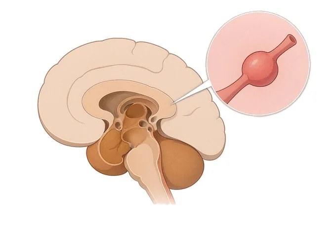
A cerebral aneurysm is a bulge in the wall of an artery in the brain, most often where the vessel branches. The majority never cause anything at all: they are found by chance on a scan done for an unrelated reason, and most of them will never bleed. That is the first thing worth knowing, because the word is frightening out of proportion to what most of them do. The second is that the small proportion that do bleed do so catastrophically, and on a different clock. An unruptured aneurysm is a decision to be made over weeks, weighing the risk of the aneurysm against the risk of the treatment. A ruptured one is an emergency in which the same decision has to be made within hours, because bleeding a second time is the single thing most likely to make the outcome worse.
-
National
Stroke Center construction unit, and a national GCP base
-
3D DSA
Alongside 3.0T MRI, dual-source CT, PET-CT and 24-hour video EEG
-
48 hours
Written second-opinion report after complete records arrive
-
124+
Countries and regions our international patients come from
What an aneurysm is, and the two clocks it runs on
An artery in the brain is built to carry blood under pressure for a lifetime. Where the vessel branches, the wall is structurally weaker than it is along the straight sections, and it is there that a bulge most often forms. That bulge is an aneurysm: not a tumour, not a blockage, and not a growth in any ordinary sense — a local failure of the wall, which enlarges slowly and silently over years.
The first thing to say about it is that it is common and usually quiet. Imaging and autopsy series find unruptured aneurysms in a small percentage of adults, commonly quoted at around two to three per cent, and most of those will never bleed. A discovery of this kind is therefore not a diagnosis that demands an immediate response; it is a finding that has to be placed on a scale, and the scale is built from the size, the site and the shape.
The second thing is what makes the condition dangerous, and it is a matter of where the blood goes. An aneurysm sits in the subarachnoid space — the compartment between the brain and the skull that is filled with cerebrospinal fluid. When it bleeds, blood escapes into that space under arterial pressure, and the result is a subarachnoid haemorrhage rather than a bleed inside the brain tissue. That distinction explains the symptoms: a bleed into the tissue produces a deficit such as weakness or loss of speech, while a bleed into the space produces a headache, vomiting, neck stiffness and a fall in consciousness.
It also explains the two clocks. Before a bleed, the question is whether to treat at all, and it can be answered over weeks. After a bleed, the aneurysm has to be sealed, and the reason is specific: bleeding again is most likely in the first day, and a second bleed is far worse than the first. The same decision, made on a completely different timescale.
Shape, size and site — the three things that decide
An aneurysm is not described by one feature. Three are read together, and between them they determine both the risk of bleeding and which of the two treatments is even possible.
Shape comes first because it carries the mechanism. The common form is saccular — often called a berry aneurysm — a rounded pouch arising from one side of the vessel at a branch point, with a neck where it joins the parent artery. Less common forms behave differently: a fusiform aneurysm widens the whole circumference of the vessel rather than pouching out of one side, and it often cannot be clipped or coiled in the ordinary way; a dissecting aneurysm follows a tear inside the wall; and an infective aneurysm arises where an infection has damaged the wall.
Size is the number most patients are given, and it matters because the risk of rupture rises with it. Aneurysms are conventionally grouped as small (under about 7 mm), medium (roughly 7–12 mm), large (13–24 mm) and giant (25 mm or more). The boundary that appears most often in the literature is 7 mm, and it is the reason a small aneurysm in a low-risk position is frequently watched rather than treated. Size is not the whole answer, however: a substantial share of aneurysms that bleed are small, so a small measurement reduces risk rather than removing it.
Site matters for two separate reasons. It changes the risk: aneurysms on the posterior circulation — the vertebrobasilar system, including the tip of the basilar artery — are reported to bleed more often at a given size than those at the front of the circle of Willis. And it changes what can be done: an aneurysm at the bifurcation of the middle cerebral artery is surgically accessible in a way that one at the top of the basilar artery is not, while an aneurysm whose neck incorporates a branch that must be preserved may favour a technique that does not require the neck to be closed.
Size, site and whether it has bled
An aneurysm is assessed along several axes at once. The ones that decide what happens are not all anatomical: the single largest division is whether it has bled, because that changes the timescale on which every other judgement has to be made.
| Axis | How it is described |
|---|---|
| Whether it has bled | Unruptured, or ruptured. This is the axis that sets the clock: unruptured aneurysms allow a decision made over weeks with the patient's own priorities weighed in, while a ruptured aneurysm has to be sealed within the first day because a second bleed is most likely in that window. |
| Size | Grouped as small (under about 7 mm), medium (7–12 mm), large (13–24 mm) and giant (25 mm or more), measured on angiography. The risk of rupture rises with size, and 7 mm is the boundary most often used when deciding between watching and treating — but a substantial share of bleeds come from aneurysms below it. |
| Site | Anterior communicating artery, posterior communicating artery, middle cerebral artery bifurcation, the cavernous segment of the internal carotid, and the posterior circulation including the basilar tip. Posterior circulation aneurysms are reported to bleed more often at a given size, and site also decides which approach is anatomically possible. |
| Neck and shape | A narrow neck favours straightforward coiling; a wide neck, or one that incorporates a branch that must be preserved, may require a stent, a flow diverter or a clip. Irregularity of the dome, and a small additional outpouching, are features noted on angiography because they are associated with rupture. |
| Growth and multiplicity | Whether the aneurysm has enlarged on interval imaging, and whether there is more than one. Growth shifts the balance towards treatment; multiple aneurysms require the one that bled to be identified before anything is sealed. |
| Clinical grade after a bleed | The level of consciousness and the neurological deficit, recorded on a standard scale and repeated. It is the strongest guide to outcome after a rupture, and it is reassessed rather than recorded once, because a fall in it is treated as an emergency. |
| Time since the bleed, and the vasospasm window | How many days have passed. Rebleeding dominates the first day; narrowing of the arteries — vasospasm, which can cause delayed deterioration — is watched for over the days that follow and is the reason monitoring continues after the aneurysm has been secured. This is the axis that governs the length and the shape of the stay. |
Read together, these axes do not answer "how dangerous is this aneurysm". They answer which of three situations this is: a finding that should be watched with interval imaging and the risk factors addressed; an unruptured aneurysm in which the balance of risks favours sealing it now; or a ruptured aneurysm in which the decision has a deadline measured in hours.
How an aneurysm is found and described
The sequence depends on which clock is running. After a suspected bleed the order is fixed and urgent; before one, the question is which study gives the necessary detail with the least burden.
| Investigation | What it settles |
|---|---|
| Non-contrast CT of the head | Whether blood has reached the subarachnoid space. Fresh blood appears bright in the cisterns and along the surface of the brain, and the scan is most reliable in the first hours — its sensitivity falls with time, which is why a normal scan later in the day does not end the assessment. The same scan shows hydrocephalus, blood in the ventricles, and any bleed into the tissue itself. |
| CT angiography | Whether there is an aneurysm, where it is, and how it is shaped. It gives the size, the width of the neck, the relationship to neighbouring branches and the presence of more than one — enough in most cases to plan treatment, and obtainable in minutes. |
| MRI and MR angiography | Used mainly for an aneurysm that has not bled: to detect one in a patient being screened, and to follow one that is being watched. It involves no ionising radiation, which is part of why it is preferred for repeated interval imaging. |
| Digital subtraction angiography | The reference study when treatment is being planned. It shows the aneurysm filling in real time, demonstrates the branches that arise from or near the neck, and is what the decision between coiling and clipping is usually made on. It is performed by the team that would carry out the treatment. |
| Lumbar puncture | Reserved for a convincing story with a normal CT. Blood that does not clear between the first and last tubes of fluid is the finding that matters, and the test is meaningful only after enough hours have passed for the scan to have become less reliable. |
A report worth having therefore answers five things rather than one: is there an aneurysm, where is it, how big is it, what does the neck look like, and is there more than one — and, after a bleed, what the blood on the first scan says about the risk of the days to come.
Why patients travel to China for aneurysm treatment
A patient with a suspected rupture needs the nearest hospital able to treat it, and no one should travel during one. Travel becomes reasonable afterwards, and in three situations: when an unruptured aneurysm has been found and the decision between watching, coiling and clipping needs a second opinion; when an aneurysm has been secured and the question is what follow-up imaging should look like; and when the work ahead is rehabilitation after a bleed.
Our neurology and neurosurgery services are organised as a standardised stroke centre built to national stroke-centre standards, and are a National Stroke Center construction unit and national GCP base. Cerebrovascular cases are planned jointly by neurology, neurosurgery and rehabilitation rather than handed from one department to the next, and interventional work — the treatment of aneurysms, arteriovenous malformations, Moyamoya disease and arterial stenosis — is carried out on 3D DSA, alongside 3.0T MRI, dual-source CT, PET-CT and 24-hour video EEG.
The practical questions are answered in the same way as for any other condition we treat. Records reviewed by a neurovascular specialist receive a written opinion within about 48 hours of complete records arriving, and the opinion states what the images show, whether the aneurysm is suitable for coiling or for clipping or for both, and what remains uncertain. Complete treatment in China typically runs at 30–50% of comparable care in Europe or the United States. We work with more than 30 commercial insurers for direct billing and have received patients from 124 countries and regions. Where the question is whether a small unruptured aneurysm should be watched or sealed, that is often answerable from images alone, and a video consultation is a reasonable first step rather than a journey.










